Strohmeier Karin, Hofmann Martina, Jacak Jaroslaw, Narzt Marie-Sophie, Wahlmueller Marlene, Mairhofer Mario, Schaedl Barbara, Holnthoner Wolfgang, Barsch Martin, Sandhofer Matthias, Wolbank Susanne, Priglinger Eleni
Ludwig Boltzmann Institute for Traumatology in Cooperation with the AUVA, 1200 Vienna, Austria.
Austrian Cluster for Tissue Regeneration, 1200 Vienna, Austria.
Biomedicines. 2022 May 18;10(5):1163. doi: 10.3390/biomedicines10051163.
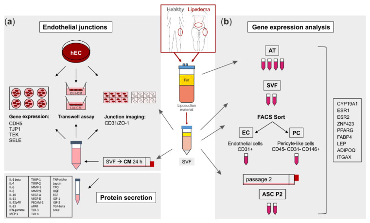
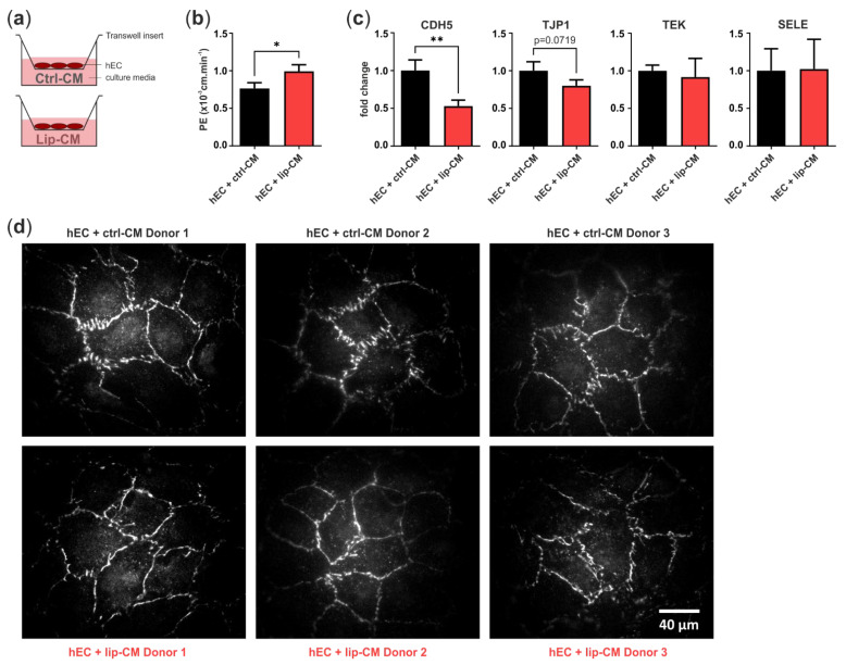
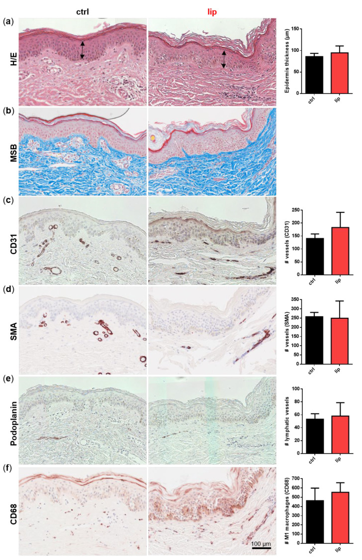
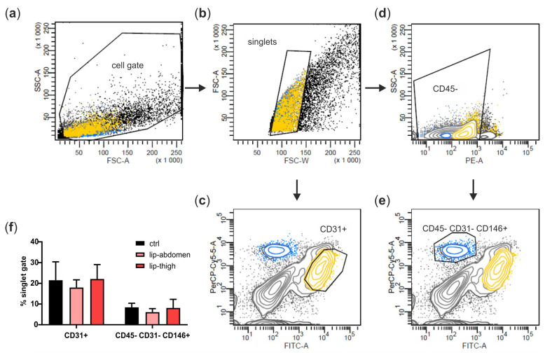
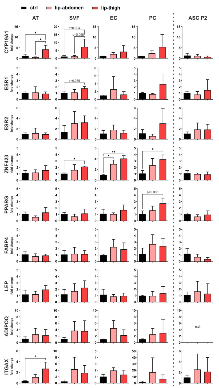
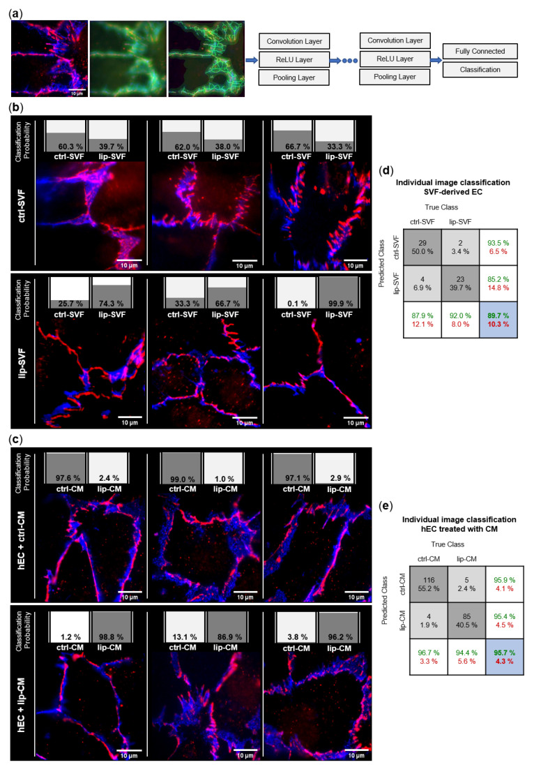
Lipedema is a chronic, progressive disease of adipose tissue with unknown etiology. Based on the relevance of the stromal vascular fraction (SVF) cell population in lipedema, we performed a thorough characterization of subcutaneous adipose tissue, SVF isolated thereof and the sorted populations of endothelial cells (EC), pericytes and cultured adipose-derived stromal/stem cells (ASC) of early-stage lipedema patients. We employed histological and gene expression analysis and investigated the endothelial barrier by immunofluorescence and analysis of endothelial permeability in vitro. Although there were no significant differences in histological stainings, we found altered gene expression of factors relevant for local estrogen metabolism (aromatase), preadipocyte commitment (ZNF423) and immune cell infiltration (CD11c) in lipedema on the tissue level, as well as in distinct cellular subpopulations. Machine learning analysis of immunofluorescence images of CD31 and ZO-1 revealed a morphological difference in the cellular junctions of EC cultures derived from healthy and lipedema individuals. Furthermore, the secretome of lipedema-derived SVF cells was sufficient to significantly increase leakiness of healthy human primary EC, which was also reflected by decreased mRNA expression of VE-cadherin. Here, we showed for the first time that the secretome of SVF cells creates an environment that triggers endothelial barrier dysfunction in early-stage lipedema. Moreover, since alterations in gene expression were detected on the cellular and/or tissue level, the choice of sample material is of high importance in elucidating this complex disease.
脂肪性水肿是一种病因不明的慢性、进行性脂肪组织疾病。基于脂肪性水肿中基质血管成分(SVF)细胞群的相关性,我们对早期脂肪性水肿患者的皮下脂肪组织、从中分离的SVF以及分选的内皮细胞(EC)、周细胞和培养的脂肪来源的基质/干细胞(ASC)群体进行了全面表征。我们采用了组织学和基因表达分析,并通过免疫荧光和体外内皮通透性分析来研究内皮屏障。尽管组织学染色没有显著差异,但我们发现在组织水平以及不同细胞亚群中,与局部雌激素代谢(芳香化酶)、前脂肪细胞定向(ZNF423)和免疫细胞浸润(CD11c)相关的因子的基因表达发生了改变。对CD31和ZO-1免疫荧光图像的机器学习分析揭示了源自健康个体和脂肪性水肿个体的EC培养物细胞连接的形态差异。此外,脂肪性水肿来源的SVF细胞的分泌组足以显著增加健康人原代EC的通透性,这也反映在VE-钙黏蛋白mRNA表达的降低上。在这里,我们首次表明SVF细胞的分泌组创造了一种环境,在早期脂肪性水肿中引发内皮屏障功能障碍。此外,由于在细胞和/或组织水平上检测到基因表达的改变,在阐明这种复杂疾病时,样本材料的选择至关重要。