Kunze Philipp, Kreiss Lucas, Novosadová Vendula, Roehe Adriana V, Steinmann Sara, Prochazka Jan, Geppert Carol I, Hartmann Arndt, Schürmann Sebastian, Friedrich Oliver, Schneider-Stock Regine
Experimental Tumor Pathology, Institute of Pathology, University Hospital Erlangen, Friedrich-Alexander University Erlangen-Nürnberg (FAU), 91054 Erlangen, Germany.
Institute of Medical Biotechnology, Friedrich-Alexander University Erlangen-Nürnberg (FAU), Paul-Gordan-Str. 3, 91052 Erlangen, Germany.
Cancers (Basel). 2022 May 10;14(10):2364. doi: 10.3390/cancers14102364.
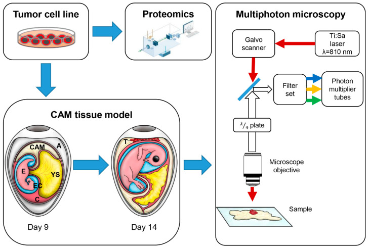
Cancer cells facilitate tumor growth by creating favorable tumor micro-environments (TME), altering homeostasis and immune response in the extracellular matrix (ECM) of surrounding tissue. A potential factor that contributes to TME generation and ECM remodeling is the cytoskeleton-associated human death-associated protein kinase 1 (DAPK1). Increased tumor cell motility and de-adhesion (thus, promoting metastasis), as well as upregulated plasminogen-signaling, are shown when functionally analyzing the DAPK1 ko-related proteome. However, the systematic investigation of how tumor cells actively modulate the ECM at the tissue level is experimentally challenging since animal models do not allow direct experimental access while artificial in vitro scaffolds cannot simulate the entire complexity of tissue systems. Here, we used the chorioallantoic membrane (CAM) assay as a natural, collagen-rich tissue model in combination with all-optical experimental access by multiphoton microscopy (MPM) to study the ECM remodeling potential of colorectal tumor cells with and without DAPK1 in situ and even in vivo. This approach demonstrates the suitability of the CAM assay in combination with multiphoton microscopy for studying collagen remodeling during tumor growth. Our results indicate the high ECM remodeling potential of DAPK1 ko tumor cells at the tissue level and support our findings from proteomics.
癌细胞通过创造有利的肿瘤微环境(TME)促进肿瘤生长,改变周围组织细胞外基质(ECM)中的稳态和免疫反应。细胞骨架相关的人类死亡相关蛋白激酶1(DAPK1)是促成TME生成和ECM重塑的一个潜在因素。在对与DAPK1基因敲除相关的蛋白质组进行功能分析时,发现肿瘤细胞的运动性和去黏附能力增强(从而促进转移),同时纤溶酶原信号上调。然而,在组织水平上系统研究肿瘤细胞如何主动调节ECM在实验上具有挑战性,因为动物模型不允许直接进行实验,而人工体外支架无法模拟组织系统的全部复杂性。在这里,我们使用绒毛尿囊膜(CAM)试验作为一种天然的、富含胶原蛋白的组织模型,并结合多光子显微镜(MPM)的全光学实验方法,来原位甚至在体内研究有无DAPK1的结直肠肿瘤细胞的ECM重塑潜力。这种方法证明了CAM试验结合多光子显微镜在研究肿瘤生长过程中胶原蛋白重塑方面的适用性。我们的结果表明,DAPK1基因敲除肿瘤细胞在组织水平上具有很高的ECM重塑潜力,并支持了我们蛋白质组学的研究结果。