Yokosuka Kimiaki, Sato Kimiaki, Yamada Kei, Yoshida Tatsuhiro, Shimazaki Takahiro, Morito Shinji, Nishida Kouta, Matsuo Atsushi, Fudo Takuma, Shiba Naoto
Department of Orthopedic Surgery, Kurume University School of Medicine, 67 Asahi-machi, Kurume-shi 830-0011, Japan.
Diagnostics (Basel). 2022 May 19;12(5):1267. doi: 10.3390/diagnostics12051267.
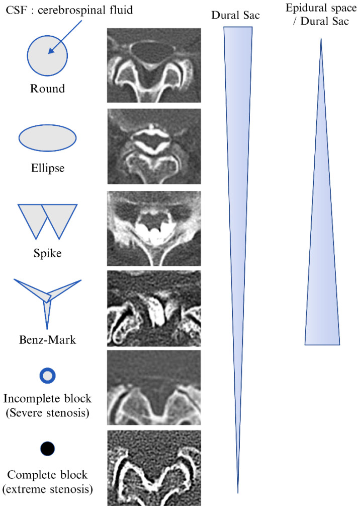
This study was conducted to analyze the findings and benefits of computed tomography (CT) epidurography in patients with low back and leg pain and compare these findings with those of magnetic resonance imaging (MRI) images. In total, 495 intervertebral discs from 99 patients with low back and leg pain who underwent percutaneous epidural adhesiolysis (epidural neuroplasty or percutaneous adhesiolysis) were examined. The axial views of CT epidurography were classified into six types to examine each intervertebral disc: round type, ellipse type, spike type, Benz mark, incomplete block, complete block, and non-contrast. MRI images were graded from A to D using the Schizas classification. Notably, 176 images were round-type and ellipse-type axial views, and 138 were spike-type and Benz-mark views; Schizas classification Grades A and B were observed in 272 and 47 MRI images, respectively. The incomplete block and complete block axial images did not significantly differ in CT epidurography and Schizas classification Grades C and D. The images showing Benz marks existed only at the L4/5 and L5/S intervertebral levels and only in 14.7% of patients. The ratio of normal shadows differed between MRI images and CT epidurography. Therefore, CT epidurography may enable a detailed evaluation of the epidural space.
本研究旨在分析计算机断层扫描(CT)脊髓造影在腰腿痛患者中的检查结果及益处,并将这些结果与磁共振成像(MRI)图像的结果进行比较。总共检查了99例接受经皮硬膜外粘连松解术(硬膜外神经成形术或经皮粘连松解术)的腰腿痛患者的495个椎间盘。CT脊髓造影的轴向视图分为六种类型以检查每个椎间盘:圆形、椭圆形、尖峰型、本茨氏征、不完全阻塞、完全阻塞和无造影剂。MRI图像使用Schizas分类从A到D进行分级。值得注意的是,176幅图像为圆形和椭圆形轴向视图,138幅为尖峰型和本茨氏征视图;Schizas分类的A级和B级分别在272幅和47幅MRI图像中观察到。CT脊髓造影中的不完全阻塞和完全阻塞轴向图像与Schizas分类的C级和D级没有显著差异。显示本茨氏征的图像仅存在于L4/5和L5/S椎间盘水平,且仅在14.7%的患者中出现。MRI图像和CT脊髓造影中正常阴影的比例不同。因此,CT脊髓造影可能有助于对硬膜外间隙进行详细评估。