• 文献检索
  • 文档翻译
  • 深度研究
  • 学术资讯
  • Suppr Zotero 插件Zotero 插件
  • 邀请有礼
  • 套餐&价格
  • 历史记录
应用&插件
Suppr Zotero 插件Zotero 插件浏览器插件Mac 客户端Windows 客户端微信小程序
定价
高级版会员购买积分包购买API积分包
服务
文献检索文档翻译深度研究API 文档MCP 服务
关于我们
关于 Suppr公司介绍联系我们用户协议隐私条款
关注我们

Suppr 超能文献

核心技术专利:CN118964589B侵权必究
粤ICP备2023148730 号-1Suppr @ 2026

文献检索

告别复杂PubMed语法,用中文像聊天一样搜索,搜遍4000万医学文献。AI智能推荐,让科研检索更轻松。

立即免费搜索

文件翻译

保留排版,准确专业,支持PDF/Word/PPT等文件格式,支持 12+语言互译。

免费翻译文档

深度研究

AI帮你快速写综述,25分钟生成高质量综述,智能提取关键信息,辅助科研写作。

立即免费体验

鉴定永生化人小胶质细胞系作为研究糖尿病视网膜病变的工具。

Characterization of an Immortalized Human Microglial Cell Line as a Tool for the Study of Diabetic Retinopathy.

机构信息

Department of Medical Sciences, University of Torino, 10126 Torino, Italy.

出版信息

Int J Mol Sci. 2022 May 20;23(10):5745. doi: 10.3390/ijms23105745.

DOI:10.3390/ijms23105745
PMID:35628555
原文链接:https://pmc.ncbi.nlm.nih.gov/articles/PMC9145666/
Abstract

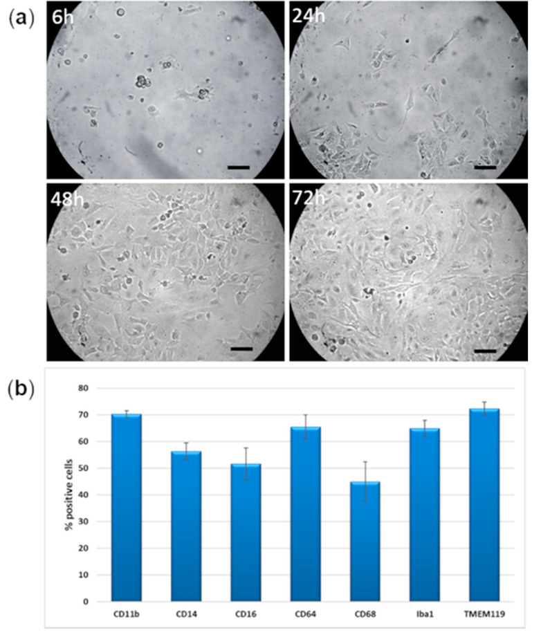
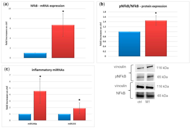
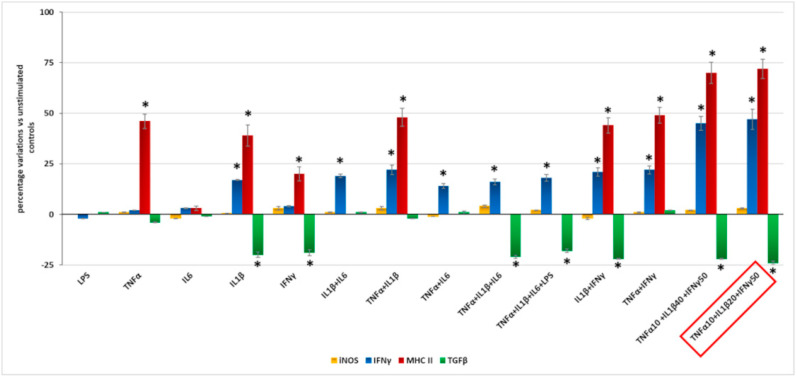
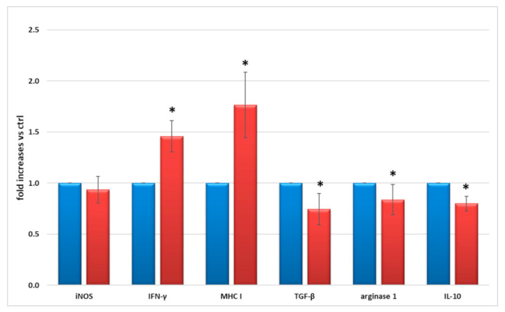
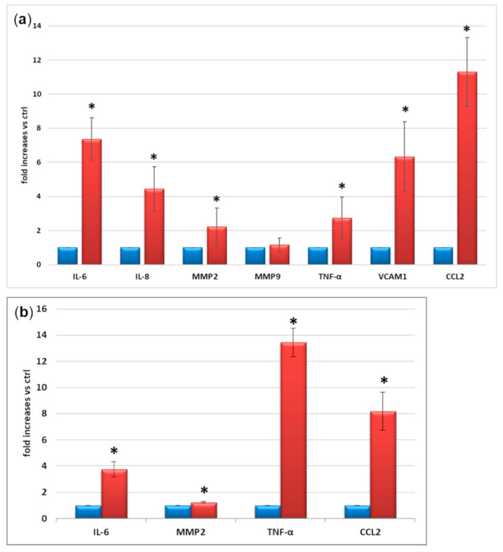
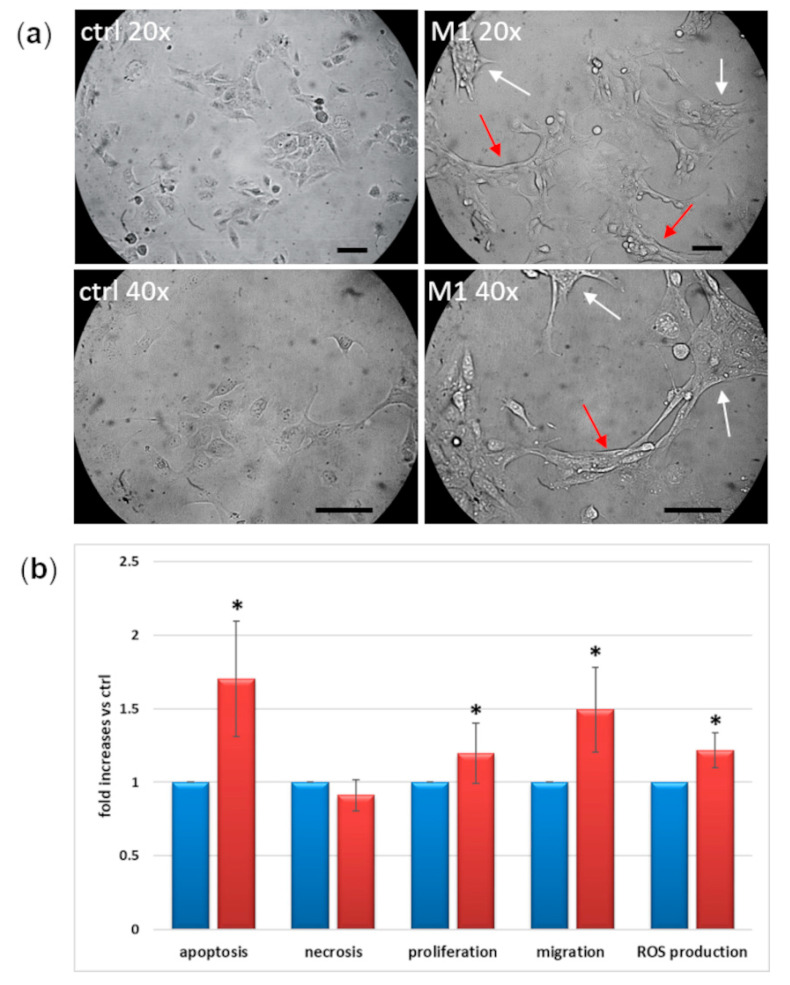
The complexity of the retinal structure reflects on the difficulty to describe its composite cell interactions. Microglia is responsible for the immune reaction to inflammatory stimuli during diabetic retinopathy (DR), but most studies still use rodent cells. We characterized a commercially available immortalized human microglial line and tested its susceptibility to inflammation, to study the interactions between the neuro-vascular retinal portions in species-specific models. After checking the expression of microglial markers, we tried lipopolysaccharide (LPS) stimulation and several pro-inflammatory cocktails to select the best combination able to induce a significant M1 (inflammatory) response. We measured M1 induction through the expression of pro- and anti-inflammatory molecules and performed morphologic and functional assays. Marker expression confirmed the human microglial derivation of these cells. Differently from rodents, LPS did not induce a M1 profile. The best pro-inflammatory stimulus was an interleukin-1β + tumor necrosis factor-α + interferon-γ cocktail, which induced morphology changes and increased proliferation, apoptosis, migration, reactive oxygen species, and the expression of inflammatory cytokines and miRNAs. In conclusion, this microglial line proved potentially useful to investigate the cascade of events leading to DR. In perspective, co-culture models involving microvascular cells will help in the understanding of multifaceted interactions of the neurovascular unit.

摘要

视网膜结构的复杂性反映在描述其复合细胞相互作用的难度上。小胶质细胞负责糖尿病视网膜病变 (DR) 中对炎症刺激的免疫反应,但大多数研究仍使用啮齿动物细胞。我们对一种可商购的永生化人小胶质细胞系进行了特征描述,并测试了其对炎症的敏感性,以在种特异性模型中研究神经血管视网膜部分之间的相互作用。在检查了小胶质细胞标志物的表达后,我们尝试了脂多糖 (LPS) 刺激和几种促炎鸡尾酒,以选择能够诱导显著 M1(炎症)反应的最佳组合。我们通过表达促炎和抗炎分子来测量 M1 诱导,并进行形态和功能测定。标志物表达证实了这些细胞来源于人类小胶质细胞。与啮齿动物不同,LPS 不会诱导 M1 特征。最佳的促炎刺激是白细胞介素 1β+肿瘤坏死因子-α+干扰素-γ 鸡尾酒,它诱导形态变化并增加增殖、凋亡、迁移、活性氧和炎症细胞因子和 miRNA 的表达。总之,这条小胶质细胞系被证明对研究导致 DR 的一系列事件具有潜在的用处。展望未来,涉及微血管细胞的共培养模型将有助于理解神经血管单元的多方面相互作用。

https://cdn.ncbi.nlm.nih.gov/pmc/blobs/d087/9145666/e96b363e8266/ijms-23-05745-g006.jpg
https://cdn.ncbi.nlm.nih.gov/pmc/blobs/d087/9145666/25e37a90af2a/ijms-23-05745-g001.jpg
https://cdn.ncbi.nlm.nih.gov/pmc/blobs/d087/9145666/961751b38cb5/ijms-23-05745-g002.jpg
https://cdn.ncbi.nlm.nih.gov/pmc/blobs/d087/9145666/4b54733c4070/ijms-23-05745-g003.jpg
https://cdn.ncbi.nlm.nih.gov/pmc/blobs/d087/9145666/184ffa0c55ff/ijms-23-05745-g004.jpg
https://cdn.ncbi.nlm.nih.gov/pmc/blobs/d087/9145666/73fcd7860f30/ijms-23-05745-g005.jpg
https://cdn.ncbi.nlm.nih.gov/pmc/blobs/d087/9145666/e96b363e8266/ijms-23-05745-g006.jpg

相似文献

1
Characterization of an Immortalized Human Microglial Cell Line as a Tool for the Study of Diabetic Retinopathy.鉴定永生化人小胶质细胞系作为研究糖尿病视网膜病变的工具。
Int J Mol Sci. 2022 May 20;23(10):5745. doi: 10.3390/ijms23105745.
2
Release of Pro-Inflammatory/Angiogenic Factors by Retinal Microvascular Cells Is Mediated by Extracellular Vesicles Derived from M1-Activated Microglia.M1 活化小胶质细胞来源的细胞外囊泡介导视网膜微血管细胞促炎/血管生成因子的释放。
Int J Mol Sci. 2023 Dec 19;25(1):15. doi: 10.3390/ijms25010015.
3
Pro-inflammatory cytokines and lipopolysaccharide induce changes in cell morphology, and upregulation of ERK1/2, iNOS and sPLA₂-IIA expression in astrocytes and microglia.促炎细胞因子和脂多糖诱导星形胶质细胞和小胶质细胞形态改变,并上调 ERK1/2、iNOS 和 sPLA₂-IIA 的表达。
J Neuroinflammation. 2011 Sep 24;8:121. doi: 10.1186/1742-2094-8-121.
4
Crocin Inhibits Oxidative Stress and Pro-inflammatory Response of Microglial Cells Associated with Diabetic Retinopathy Through the Activation of PI3K/Akt Signaling Pathway.西红花苷通过激活PI3K/Akt信号通路抑制与糖尿病视网膜病变相关的小胶质细胞的氧化应激和促炎反应。
J Mol Neurosci. 2017 Apr;61(4):581-589. doi: 10.1007/s12031-017-0899-8. Epub 2017 Feb 25.
5
Minocycline inhibits LPS-induced retinal microglia activation.米诺环素抑制脂多糖诱导的视网膜小胶质细胞活化。
Neurochem Int. 2005 Jul;47(1-2):152-8. doi: 10.1016/j.neuint.2005.04.018.
6
Characterization of a functionally active primary microglial cell culture from the pig retina.猪视网膜原代小胶质细胞功能活性培养的特征。
Exp Eye Res. 2019 Aug;185:107670. doi: 10.1016/j.exer.2019.05.010. Epub 2019 May 16.
7
Baicalein reduces inflammatory process in a rodent model of diabetic retinopathy.黄芩素可减轻糖尿病性视网膜病变啮齿动物模型中的炎症过程。
Invest Ophthalmol Vis Sci. 2009 May;50(5):2319-27. doi: 10.1167/iovs.08-2642. Epub 2008 Nov 14.
8
Anti-Inflammatory (M2) Response Is Induced by a sp-Iminosugar Glycolipid Sulfoxide in Diabetic Retinopathy.抗炎(M2)反应是由糖尿病视网膜病变中的 sp-亚氨基糖脂硫氧化物诱导的。
Front Immunol. 2021 Mar 18;12:632132. doi: 10.3389/fimmu.2021.632132. eCollection 2021.
9
Cyclic AMP is a key regulator of M1 to M2a phenotypic conversion of microglia in the presence of Th2 cytokines.在存在Th2细胞因子的情况下,环磷酸腺苷(Cyclic AMP)是小胶质细胞从M1型向M2a型表型转化的关键调节因子。
J Neuroinflammation. 2016 Jan 13;13:9. doi: 10.1186/s12974-015-0463-9.
10
Melatonin Maintains Inner Blood-Retinal Barrier by Regulating Microglia Inhibition of PI3K/Akt/Stat3/NF-κB Signaling Pathways in Experimental Diabetic Retinopathy.褪黑素通过调节小胶质细胞抑制实验性糖尿病视网膜病变中 PI3K/Akt/Stat3/NF-κB 信号通路来维持血视网膜内屏障。
Front Immunol. 2022 Mar 15;13:831660. doi: 10.3389/fimmu.2022.831660. eCollection 2022.

引用本文的文献

1
Involvement of lncRNAs NEAT1 and ZBTB11-AS1 in Active and Persistent HIV-1 Infection in C20 Human Microglial Cell Line.lncRNAs NEAT1和ZBTB11-AS1在C20人小胶质细胞系中参与HIV-1的活跃和持续感染
Int J Mol Sci. 2025 May 15;26(10):4745. doi: 10.3390/ijms26104745.
2
Human Microglia-Like Cells Differentiated from Monocytes with GM-CSF and IL-34 Show Phagocytosis of α-Synuclein Aggregates and C/EBPβ-Dependent Proinflammatory Activation.用粒细胞-巨噬细胞集落刺激因子(GM-CSF)和白细胞介素-34(IL-34)从单核细胞分化而来的人小胶质细胞样细胞表现出对α-突触核蛋白聚集体的吞噬作用以及C/EBPβ依赖性促炎激活。
Mol Neurobiol. 2025 Jan;62(1):756-772. doi: 10.1007/s12035-024-04289-z. Epub 2024 Jun 20.
3

本文引用的文献

1
Microglia and Inflammatory Responses in Diabetic Retinopathy.小胶质细胞与糖尿病视网膜病变中的炎症反应。
Front Immunol. 2020 Nov 6;11:564077. doi: 10.3389/fimmu.2020.564077. eCollection 2020.
2
IgG Immune Complexes Break Immune Tolerance of Human Microglia.IgG 免疫复合物打破人类小神经胶质细胞的免疫耐受。
J Immunol. 2020 Nov 1;205(9):2511-2518. doi: 10.4049/jimmunol.2000130. Epub 2020 Sep 23.
3
Sulforaphane Inhibits MGO-AGE-Mediated Neuroinflammation by Suppressing NF-κB, MAPK, and AGE-RAGE Signaling Pathways in Microglial Cells.
Chikungunya virus infection in human microglial C20 cells induces mitochondria-mediated apoptosis.
人源小胶质细胞 C20 中感染基孔肯雅病毒诱导线粒体介导的细胞凋亡。
Front Cell Infect Microbiol. 2024 Apr 23;14:1380736. doi: 10.3389/fcimb.2024.1380736. eCollection 2024.
4
Release of Pro-Inflammatory/Angiogenic Factors by Retinal Microvascular Cells Is Mediated by Extracellular Vesicles Derived from M1-Activated Microglia.M1 活化小胶质细胞来源的细胞外囊泡介导视网膜微血管细胞促炎/血管生成因子的释放。
Int J Mol Sci. 2023 Dec 19;25(1):15. doi: 10.3390/ijms25010015.
5
Downregulation of PIK3IP1 in retinal microglia promotes retinal pathological neovascularization via PI3K-AKT pathway activation.PIK3IP1 在视网膜小胶质细胞中的下调通过 PI3K-AKT 通路的激活促进视网膜病理性血管新生。
Sci Rep. 2023 Aug 7;13(1):12754. doi: 10.1038/s41598-023-39473-z.
6
Assaying Microglia Functions In Vitro.体外分析小胶质细胞功能。
Cells. 2022 Oct 28;11(21):3414. doi: 10.3390/cells11213414.
萝卜硫素通过抑制小胶质细胞中的NF-κB、MAPK和AGE-RAGE信号通路来抑制MGO-AGE介导的神经炎症。
Antioxidants (Basel). 2020 Aug 26;9(9):792. doi: 10.3390/antiox9090792.
4
ROS Generation in Microglia: Understanding Oxidative Stress and Inflammation in Neurodegenerative Disease.小胶质细胞中活性氧的产生:理解神经退行性疾病中的氧化应激和炎症
Antioxidants (Basel). 2020 Aug 13;9(8):743. doi: 10.3390/antiox9080743.
5
The Role of TGFβ Signaling in Microglia Maturation and Activation.TGFβ 信号在小胶质细胞成熟和激活中的作用。
Trends Immunol. 2020 Sep;41(9):836-848. doi: 10.1016/j.it.2020.07.003. Epub 2020 Jul 30.
6
Loss of IL-10 Promotes Differentiation of Microglia to a M1 Phenotype.白细胞介素-10的缺失促进小胶质细胞向M1表型分化。
Front Cell Neurosci. 2019 Oct 9;13:430. doi: 10.3389/fncel.2019.00430. eCollection 2019.
7
Microglial Pro-Inflammatory and Anti-Inflammatory Phenotypes Are Modulated by Translocator Protein Activation.小胶质细胞的促炎和抗炎表型受转位蛋白激活的调节。
Int J Mol Sci. 2019 Sep 10;20(18):4467. doi: 10.3390/ijms20184467.
8
Pro-Inflammatory Activation of A New Immortalized Human Microglia Cell Line.一种新的永生化人小胶质细胞系的促炎激活
Brain Sci. 2019 May 15;9(5):111. doi: 10.3390/brainsci9050111.
9
Acute glucose fluctuation impacts microglial activity, leading to inflammatory activation or self-degradation.急性血糖波动会影响小胶质细胞的活性,导致炎症激活或自我降解。
Sci Rep. 2019 Jan 29;9(1):840. doi: 10.1038/s41598-018-37215-0.
10
Chronic hyperglycemia regulates microglia polarization through ERK5.慢性高血糖通过ERK5调节小胶质细胞极化。
Aging (Albany NY). 2019 Jan 26;11(2):697-706. doi: 10.18632/aging.101770.