Vos Annelotte, Vink Aryan, Kockelkoren Remko, Takx Richard A P, Celeng Csilla, Mali Willem P T M, Isgum Ivana, Bleys Ronald L A W, de Jong Pim A
Department of Pathology, University Medical Center Utrecht and Utrecht University, 3584 CX Utrecht, The Netherlands.
Department of Pathlogy, Meander Medical Center, 3800 BM Amersfoort, The Netherlands.
J Pers Med. 2022 Apr 29;12(5):711. doi: 10.3390/jpm12050711.
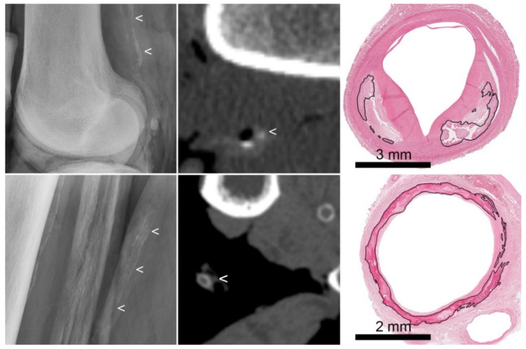
Calcifications are common in the tunica intima and tunica media of leg arteries. There is growing interest in medial arterial calcifications, as they may be modifiable with treatment. We aimed to investigate radiography and computed tomography (CT) for the detection and characterization of both types of arterial calcification in leg arteries in relation to histology. In a postmortem study we therefore investigated 24 popliteal and 24 tibial arteries. The reference standard was presence of arterial calcification and the dominance of intimal or medial calcification on histology. Radiographs and CT scans were scored for presence of calcification and for dominant intimal or medial pattern based on prespecified criteria (annularity, thickness, continuity). Both radiography and CT detected 87% of histologically proven calcifications but missed mild calcifications in 13%. When only the arteries with detected calcifications were included, a moderate agreement was observed on intimal/medial location of calcifications between histology and radiography (correct in 19/24 arteries (79%); Kappa 0.58) or CT (correct in 33/46 arterial segments (72%); Kappa 0.48). With both modalities there was a slight tendency to classify intimal calcifications as being located in the media and to miss media calcification. Our study demonstrates the potential and limitations of both radiography and CT to detect and classify arterial calcifications in leg arteries.
钙化在腿部动脉的内膜和中膜中很常见。人们对动脉中膜钙化的兴趣与日俱增,因为其可能可通过治疗得到改善。我们旨在研究X线摄影和计算机断层扫描(CT)在检测腿部动脉两种类型的动脉钙化并对其进行特征描述方面与组织学的相关性。因此,在一项尸检研究中,我们对24条腘动脉和24条胫动脉进行了研究。参考标准为动脉钙化的存在以及组织学上内膜或中膜钙化的优势情况。根据预先设定的标准(环状、厚度、连续性),对X线片和CT扫描进行钙化存在情况以及内膜或中膜主导模式的评分。X线摄影和CT均检测出了87%经组织学证实的钙化,但有13%的轻度钙化未被检测到。当仅纳入检测到钙化的动脉时,在钙化的内膜/中膜位置方面,组织学与X线摄影之间观察到中度一致性(24条动脉中有19条正确(79%);kappa值为0.58)或与CT之间(46个动脉节段中有33个正确(72%);kappa值为0.48)。两种检查方式都有将内膜钙化分类为位于中膜且漏诊中膜钙化的轻微倾向。我们的研究证明了X线摄影和CT在检测和分类腿部动脉钙化方面的潜力和局限性。