Jianu Dragos Catalin, Jianu Silviana Nina, Dan Traian Flavius, Munteanu Georgiana, Copil Alexandra, Birdac Claudiu Dumitru, Motoc Andrei Gheorghe Marius, Docu Axelerad Any, Petrica Ligia, Arnautu Sergiu Florin, Sadik Raphael, Iacob Nicoleta, Gogu Anca Elena
Department of Neurosciences-Division of Neurology, Victor Babes University of Medicine and Pharmacy, E. Murgu Sq., no.2, 300041 Timisoara, Romania.
Centre for Cognitive Research in Neuropsychiatric Pathology (NeuroPsy-Cog), Department of Neurosciences, Victor Babes University of Medicine and Pharmacy, 156 L. Rebreanu Ave., 300736 Timisoara, Romania.
Life (Basel). 2022 May 11;12(5):717. doi: 10.3390/life12050717.
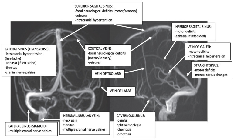
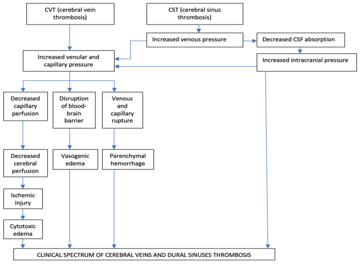
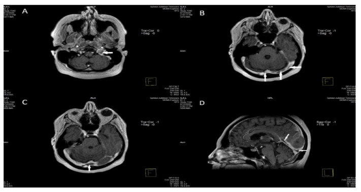
(1) Objective: This review paper aims to discuss multiple aspects of cerebral venous thrombosis (CVT), including epidemiology, etiology, pathophysiology, and clinical presentation. Different neuroimaging methods for diagnosis of CVT, such as computer tomography CT/CT Venography (CTV), and Magnetic Resonance Imaging (MRI)/MR Venography (MRV) will be presented. (2) Methods: A literature analysis using PubMed and the MEDLINE sub-engine was done using the terms: cerebral venous thrombosis, thrombophilia, and imaging. Different studies concerning risk factors, clinical picture, and imaging signs of patients with CVT were examined. (3) Results: At least one risk factor can be identified in 85% of CVT cases. Searching for a thrombophilic state should be realized for patients with CVT who present a high pretest probability of severe thrombophilia. Two pathophysiological mechanisms contribute to their highly variable clinical presentation: augmentation of venular and capillary pressure, and diminution of cerebrospinal fluid absorption. The clinical spectrum of CVT is frequently non-specific and presents a high level of clinical suspicion. Four major syndromes have been described: isolated intracranial hypertension, seizures, focal neurological abnormalities, and encephalopathy. Cavernous sinus thrombosis is the single CVT that presents a characteristic clinical syndrome. Non-enhanced CT (NECT) of the Head is the most frequently performed imaging study in the emergency department. Features of CVT on NECT can be divided into direct signs (demonstration of dense venous clot within a cerebral vein or a cerebral venous sinus), and more frequently indirect signs (such as cerebral edema, or cerebral venous infarct). CVT diagnosis is confirmed with CTV, directly detecting the venous clot as a filling defect, or MRI/MRV, which also realizes a better description of parenchymal abnormalities. (4) Conclusions: CVT is a relatively rare disorder in the general population and is frequently misdiagnosed upon initial examination. The knowledge of wide clinical aspects and imaging signs will be essential in providing a timely diagnosis.
(1) 目的:本综述旨在探讨脑静脉血栓形成(CVT)的多个方面,包括流行病学、病因、病理生理学和临床表现。还将介绍用于诊断CVT的不同神经影像学方法,如计算机断层扫描CT/CT静脉造影(CTV)和磁共振成像(MRI)/磁共振静脉造影(MRV)。(2) 方法:使用PubMed和MEDLINE子引擎进行文献分析,搜索词为:脑静脉血栓形成、血栓形成倾向和成像。研究了有关CVT患者的危险因素、临床表现和影像学征象的不同研究。(3) 结果:85%的CVT病例可识别出至少一种危险因素。对于具有严重血栓形成倾向高预测试概率的CVT患者,应进行血栓形成倾向状态的检查。两种病理生理机制导致其临床表现高度可变:小静脉和毛细血管压力升高以及脑脊液吸收减少。CVT的临床谱通常不具有特异性,临床怀疑程度高。已描述了四种主要综合征:孤立性颅内高压、癫痫发作、局灶性神经功能异常和脑病。海绵窦血栓形成是唯一呈现特征性临床综合征的CVT。头部非增强CT(NECT)是急诊科最常进行的影像学检查。NECT上CVT的特征可分为直接征象(显示脑静脉或脑静脉窦内的致密静脉血栓),更常见的是间接征象(如脑水肿或脑静脉梗死)。CTV直接将静脉血栓检测为充盈缺损,或MRI/MRV可确诊CVT,后者还能更好地描述实质异常。(4) 结论:CVT在普通人群中是一种相对罕见的疾病,初诊时经常被误诊。了解广泛的临床方面和影像学征象对于及时诊断至关重要。