• 文献检索
  • 文档翻译
  • 深度研究
  • 学术资讯
  • Suppr Zotero 插件Zotero 插件
  • 邀请有礼
  • 套餐&价格
  • 历史记录
应用&插件
Suppr Zotero 插件Zotero 插件浏览器插件Mac 客户端Windows 客户端微信小程序
定价
高级版会员购买积分包购买API积分包
服务
文献检索文档翻译深度研究API 文档MCP 服务
关于我们
关于 Suppr公司介绍联系我们用户协议隐私条款
关注我们

Suppr 超能文献

核心技术专利:CN118964589B侵权必究
粤ICP备2023148730 号-1Suppr @ 2026

文献检索

告别复杂PubMed语法,用中文像聊天一样搜索,搜遍4000万医学文献。AI智能推荐,让科研检索更轻松。

立即免费搜索

文件翻译

保留排版,准确专业,支持PDF/Word/PPT等文件格式,支持 12+语言互译。

免费翻译文档

深度研究

AI帮你快速写综述,25分钟生成高质量综述,智能提取关键信息,辅助科研写作。

立即免费体验

低氧增强外周血来源间充质基质细胞的增殖和干性。

The Proliferation and Stemness of Peripheral Blood-Derived Mesenchymal Stromal Cells Were Enhanced by Hypoxia.

机构信息

Guangzhou Institute of Traumatic Surgery, Guangzhou Red Cross Hospital, Jinan University, Guangzhou, China.

Department of Neurology, Guangzhou Red Cross Hospital, Jinan University, Guangzhou, China.

出版信息

Front Endocrinol (Lausanne). 2022 May 12;13:873662. doi: 10.3389/fendo.2022.873662. eCollection 2022.

DOI:10.3389/fendo.2022.873662
PMID:35634504
原文链接:https://pmc.ncbi.nlm.nih.gov/articles/PMC9134856/
Abstract

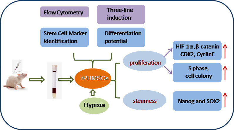
This study aimed to address the dilemma of low peripheral blood-derived mesenchymal stromal cell (PBMSC) activity and reduced phenotype in bone or cartilage tissue engineering. Rat PBMSCs (rPBMSCs) were obtained by density gradient centrifugation, and stromal cell characteristics were confirmed by flow cytometry (FCM) and multi-differentiation potential induction experiments. Cell growth curve, viability experiments, and clone formation experiments were performed by [3-(4,5-dimethylthiazol-2-yl)-5-(3-carboxymethoxyphenyl)-2-(4-sulfophenyl)-2H-tetrazolium] (MTS) and cell counting, and the cell cycle was confirmed by cell FCM. The proliferation signal pathway and stemness-related proteins were detected by molecular methods including Western blot and real-time polymerase chain reaction. , and were highly expressed, and , and were barely expressed in rPBMSCs. rPBMSCs possessed the potential to differentiate into chondrocytes, adipocytes, and osteoblasts under their respective induction conditions. Cell growth curve and viability experiments were performed under hypoxic conditions: 19% O, 5% O, and 1% O. Specifically, 5% O accelerated the proliferation and expression of the stemness of PBMSCs. Cycle experiments proved that hypoxia promoted the cell transition from the G1 phase to the S phase. Molecular experiments confirmed that 5% O hypoxia significantly elevated the expressions of hypoxia-inducible factor 1α and β-catenin and simultaneously the expressions of cycle-related genes including and stemness-related genes including and . The appropriate concentration of hypoxia (i.e., 5% O) enhanced the proliferation and stemness of rPBMSCs and increased the multidirectional differentiation potential of stromal cells. The proposed culture method could improve the viability and maintain the phenotype of rPBMSCs in cartilage or bone tissue engineering.

摘要

本研究旨在解决外周血源性间充质基质细胞(PBMSC)活性低和在骨或软骨组织工程中表型减少的难题。通过密度梯度离心法获取大鼠 PBMSCs(rPBMSCs),并通过流式细胞术(FCM)和多向分化潜能诱导实验确认基质细胞特征。通过[3-(4,5-二甲基噻唑-2-基)-5-(3-羧基甲氧基苯基)-2-(4-磺基苯基)-2H-四唑](MTS)和细胞计数进行细胞生长曲线、活力实验和克隆形成实验,通过细胞 FCM 确认细胞周期。通过分子方法包括 Western blot 和实时聚合酶链反应检测增殖信号通路和干细胞相关蛋白。结果显示,rPBMSCs 中高表达、几乎不表达。rPBMSCs 在各自的诱导条件下具有分化为软骨细胞、脂肪细胞和成骨细胞的潜能。在低氧条件下(19% O、5% O 和 1% O)进行细胞生长曲线和活力实验。具体来说,5% O 加速了 PBMSCs 的增殖和干性表达。周期实验证明,低氧促进了细胞从 G1 期向 S 期的过渡。分子实验证实,5% O 低氧显著上调了缺氧诱导因子 1α 和 β-连环蛋白的表达,同时上调了周期相关基因如和干细胞相关基因如和。适当的低氧浓度(即 5% O)增强了 rPBMSCs 的增殖和干性,并增加了基质细胞的多向分化潜能。所提出的培养方法可以提高 rPBMSCs 在软骨或骨组织工程中的活力并维持其表型。

https://cdn.ncbi.nlm.nih.gov/pmc/blobs/4429/9134856/78d1a209c943/fendo-13-873662-g006.jpg
https://cdn.ncbi.nlm.nih.gov/pmc/blobs/4429/9134856/e4a7da2ab6ba/fendo-13-873662-g001.jpg
https://cdn.ncbi.nlm.nih.gov/pmc/blobs/4429/9134856/41151f356f26/fendo-13-873662-g002.jpg
https://cdn.ncbi.nlm.nih.gov/pmc/blobs/4429/9134856/49fa3d7e6136/fendo-13-873662-g003.jpg
https://cdn.ncbi.nlm.nih.gov/pmc/blobs/4429/9134856/88bfd476e110/fendo-13-873662-g004.jpg
https://cdn.ncbi.nlm.nih.gov/pmc/blobs/4429/9134856/b4a5f441291b/fendo-13-873662-g005.jpg
https://cdn.ncbi.nlm.nih.gov/pmc/blobs/4429/9134856/78d1a209c943/fendo-13-873662-g006.jpg

相似文献

1
The Proliferation and Stemness of Peripheral Blood-Derived Mesenchymal Stromal Cells Were Enhanced by Hypoxia.低氧增强外周血来源间充质基质细胞的增殖和干性。
Front Endocrinol (Lausanne). 2022 May 12;13:873662. doi: 10.3389/fendo.2022.873662. eCollection 2022.
2
Hypoxia with Wharton's jelly mesenchymal stem cell coculture maintains stemness of umbilical cord blood-derived CD34 cells.缺氧条件下与华通氏胶间充质干细胞共培养维持脐带血来源 CD34 细胞的干性。
Stem Cell Res Ther. 2018 Jun 13;9(1):158. doi: 10.1186/s13287-018-0902-5.
3
[Enrichment and Biological Characteristics of Peripheral Blood-derived Mesenchymal Stem Cells in Rats].[大鼠外周血源性间充质干细胞的富集及生物学特性]
Zhongguo Shi Yan Xue Ye Xue Za Zhi. 2015 Apr;23(2):506-11. doi: 10.7534/j.issn.1009-2137.2015.02.041.
4
Hypoxia reduces the osteogenic differentiation of peripheral blood mesenchymal stem cells by upregulating Notch-1 expression.缺氧通过上调 Notch-1 表达抑制外周血间充质干细胞的成骨分化。
Connect Tissue Res. 2019 Nov;60(6):583-596. doi: 10.1080/03008207.2019.1611792. Epub 2019 May 28.
5
Hypoxia enhances buffalo adipose-derived mesenchymal stem cells proliferation, stemness, and reprogramming into induced pluripotent stem cells.缺氧增强水牛脂肪间充质干细胞的增殖、干性和重编程为诱导多能干细胞。
J Cell Physiol. 2019 Aug;234(10):17254-17268. doi: 10.1002/jcp.28342. Epub 2019 Feb 25.
6
Isolation and Characterization of Rat Mesenchymal Stem Cells Derived from Granulocyte Colony-Stimulating Factor-Mobilized Peripheral Blood.从粒细胞集落刺激因子动员的外周血中分离并鉴定大鼠间充质干细胞
Cells Tissues Organs. 2015;201(6):412-422. doi: 10.1159/000445855. Epub 2016 Jun 1.
7
The effect of intervertebral disc degenerative change on biological characteristics of nucleus pulposus mesenchymal stem cell: an study in rats.椎间盘退变对髓核间充质干细胞生物学特性的影响:一项大鼠研究
Connect Tissue Res. 2019 Jul;60(4):376-388. doi: 10.1080/03008207.2019.1570168.
8
Low oxygen atmosphere facilitates proliferation and maintains undifferentiated state of umbilical cord mesenchymal stem cells in an hypoxia inducible factor-dependent manner.低氧环境以缺氧诱导因子依赖的方式促进脐带间充质干细胞的增殖并维持其未分化状态。
Cytotherapy. 2014 Jul;16(7):881-92. doi: 10.1016/j.jcyt.2014.02.009. Epub 2014 Apr 13.
9
Prolonged exposure to hypoxic milieu improves the osteogenic potential of adipose derived stem cells.长期暴露于低氧环境可提高脂肪来源干细胞的成骨潜能。
J Cell Biochem. 2015 Jul;116(7):1442-53. doi: 10.1002/jcb.25106.
10
Proliferation and apoptosis property of mesenchymal stem cells derived from peripheral blood under the culture conditions of hypoxia and serum deprivation.低氧和血清剥夺培养条件下外周血间充质干细胞的增殖和凋亡特性。
Chin Med J (Engl). 2011 Dec;124(23):3959-67.

引用本文的文献

1
Identification and validation of hypoxia-associated genes positively correlated with in pulmonary adenocarcinoma.肺腺癌中与缺氧相关且呈正相关的基因的鉴定与验证。
Transl Cancer Res. 2025 May 30;14(5):3113-3132. doi: 10.21037/tcr-2024-2265. Epub 2025 May 27.
2
Hypoxia Promotes the In Vitro Proliferation of Buffalo Spermatogonial Cells by Increasing Lactate and H3K18la Lactylation Levels.缺氧通过提高乳酸和H3K18la乳酸化水平促进水牛精原细胞的体外增殖。
Cells. 2025 Jun 3;14(11):832. doi: 10.3390/cells14110832.
3
Bone marrow mesenchymal stem cell exosomes improve fracture union via remodeling metabolism in nonunion rat model.

本文引用的文献

1
Hypoxic Culture Maintains Cell Growth of the Primary Human Valve Interstitial Cells with Stemness.低氧培养维持具有干性的原代人心脏瓣膜间质细胞的细胞生长。
Int J Mol Sci. 2021 Sep 29;22(19):10534. doi: 10.3390/ijms221910534.
2
Genetically-programmed, mesenchymal stromal cell-laden & mechanically strong 3D bioprinted scaffolds for bone repair.用于骨修复的基因编程、负载间充质基质细胞且机械强度高的3D生物打印支架。
J Control Release. 2020 Sep 10;325:335-346. doi: 10.1016/j.jconrel.2020.06.035. Epub 2020 Jul 3.
3
Bovine peripheral blood MSCs chemotax towards inflammation and embryo implantation stimuli.
骨髓间充质干细胞外泌体通过重塑骨不连大鼠模型的代谢来促进骨折愈合。
J Orthop Surg Res. 2025 Mar 25;20(1):308. doi: 10.1186/s13018-025-05721-3.
4
Regional Gene Therapy for Bone Tissue Engineering: A Current Concepts Review.骨组织工程的区域基因治疗:当前概念综述
Bioengineering (Basel). 2025 Jan 27;12(2):120. doi: 10.3390/bioengineering12020120.
5
The issue of heterogeneity of MSC-based advanced therapy medicinal products-a review.基于间充质干细胞的先进治疗药品的异质性问题——综述
Front Cell Dev Biol. 2024 Jul 26;12:1400347. doi: 10.3389/fcell.2024.1400347. eCollection 2024.
6
Application progress of single-cell sequencing technology in mesenchymal stem cells research.单细胞测序技术在间充质干细胞研究中的应用进展
Front Cell Dev Biol. 2024 Jan 9;11:1336482. doi: 10.3389/fcell.2023.1336482. eCollection 2023.
7
Regulation Mechanisms and Maintenance Strategies of Stemness in Mesenchymal Stem Cells.间质干细胞干性的调控机制与维持策略。
Stem Cell Rev Rep. 2024 Feb;20(2):455-483. doi: 10.1007/s12015-023-10658-3. Epub 2023 Nov 27.
8
HIF-1α Regulates Bone Homeostasis and Angiogenesis, Participating in the Occurrence of Bone Metabolic Diseases.HIF-1α 调节骨稳态和血管生成,参与骨代谢疾病的发生。
Cells. 2022 Nov 10;11(22):3552. doi: 10.3390/cells11223552.
9
The Influence of Intervertebral Disc Microenvironment on the Biological Behavior of Engrafted Mesenchymal Stem Cells.椎间盘微环境对植入间充质干细胞生物学行为的影响
Stem Cells Int. 2022 Nov 7;2022:8671482. doi: 10.1155/2022/8671482. eCollection 2022.
牛外周血间充质干细胞向炎症和胚胎着床刺激趋化。
J Cell Physiol. 2021 Feb;236(2):1054-1067. doi: 10.1002/jcp.29915. Epub 2020 Jul 2.
4
Targeting Cardiac Stem Cell Senescence to Treat Cardiac Aging and Disease.靶向心脏干细胞衰老治疗心脏衰老和疾病。
Cells. 2020 Jun 26;9(6):1558. doi: 10.3390/cells9061558.
5
Effect of peripheral blood-derived mesenchymal stem cells on macrophage polarization and Th17/Treg balance .外周血源性间充质干细胞对巨噬细胞极化及Th17/Treg平衡的影响
Regen Ther. 2020 May 16;14:275-283. doi: 10.1016/j.reth.2020.03.008. eCollection 2020 Jun.
6
Exosomes from conditioned media of bone marrow-derived mesenchymal stem cells promote bone regeneration by enhancing angiogenesis.骨髓间充质干细胞条件培养基来源的外泌体通过增强血管生成促进骨再生。
PLoS One. 2019 Nov 21;14(11):e0225472. doi: 10.1371/journal.pone.0225472. eCollection 2019.
7
Hypoxia reduces the osteogenic differentiation of peripheral blood mesenchymal stem cells by upregulating Notch-1 expression.缺氧通过上调 Notch-1 表达抑制外周血间充质干细胞的成骨分化。
Connect Tissue Res. 2019 Nov;60(6):583-596. doi: 10.1080/03008207.2019.1611792. Epub 2019 May 28.
8
Hypoxia impacts human MSC response to substrate stiffness during chondrogenic differentiation.缺氧会影响人骨髓间充质干细胞在软骨分化过程中对基质硬度的反应。
Acta Biomater. 2019 Apr 15;89:73-83. doi: 10.1016/j.actbio.2019.03.002. Epub 2019 Mar 4.
9
Hypoxia-inducible factor 1-alpha (HIF-1α) as a factor mediating the relationship between obesity and heart failure with preserved ejection fraction.缺氧诱导因子 1-α(HIF-1α)作为肥胖与射血分数保留心力衰竭之间关系的中介因子。
Obes Rev. 2019 May;20(5):701-712. doi: 10.1111/obr.12828. Epub 2019 Mar 3.
10
Targeting HIF-1α signaling pathway for gastric cancer treatment.靶向缺氧诱导因子-1α信号通路用于胃癌治疗。
Pharmazie. 2019 Jan 1;74(1):3-7. doi: 10.1691/ph.2019.8674.