Institute of Life Course and Medical Sciences, University of Liverpool, Liverpool, UK.
Department of Cell and Molecular Physiology, Stritch School of Medicine, Loyola University Chicago, Maywood, Illinois, USA.
FASEB J. 2022 Jul;36(7):e22318. doi: 10.1096/fj.202002588RRR.
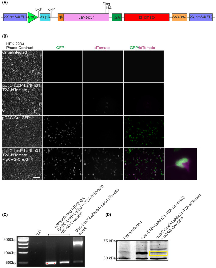
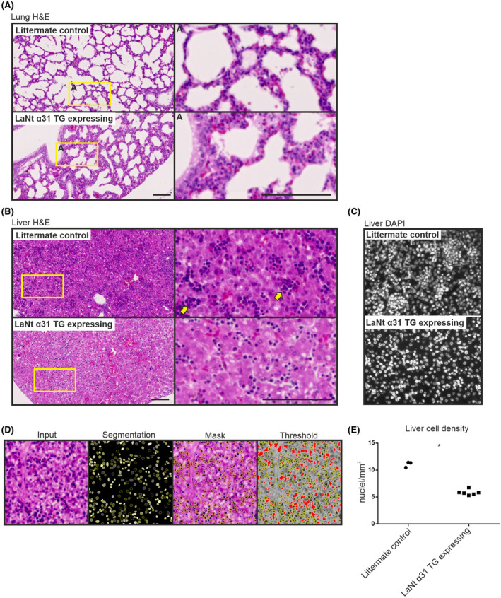
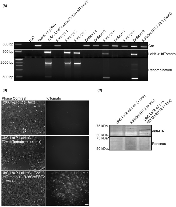
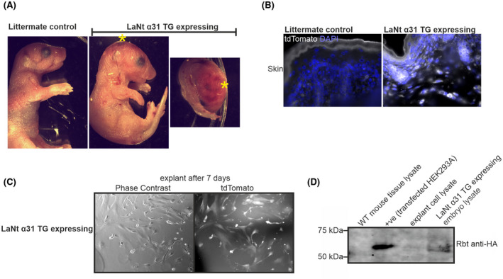
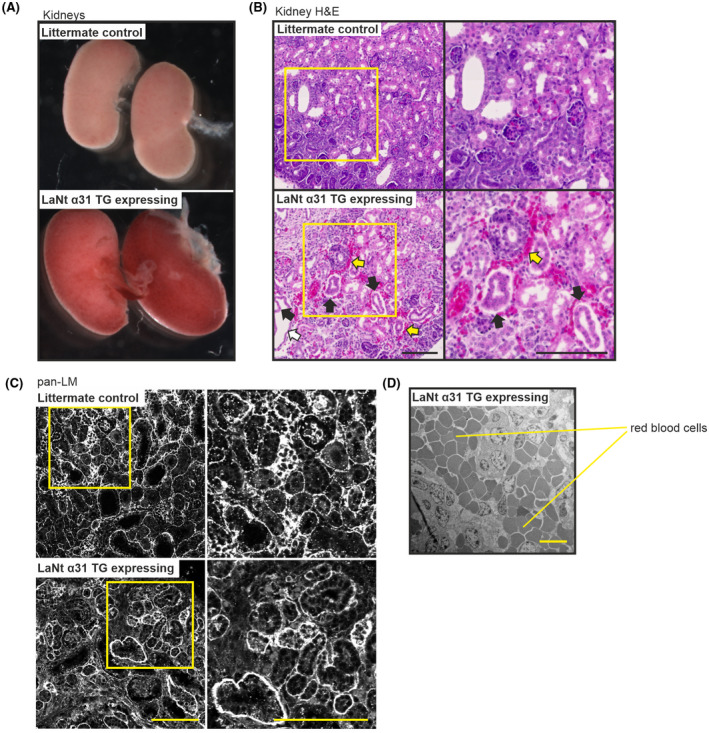
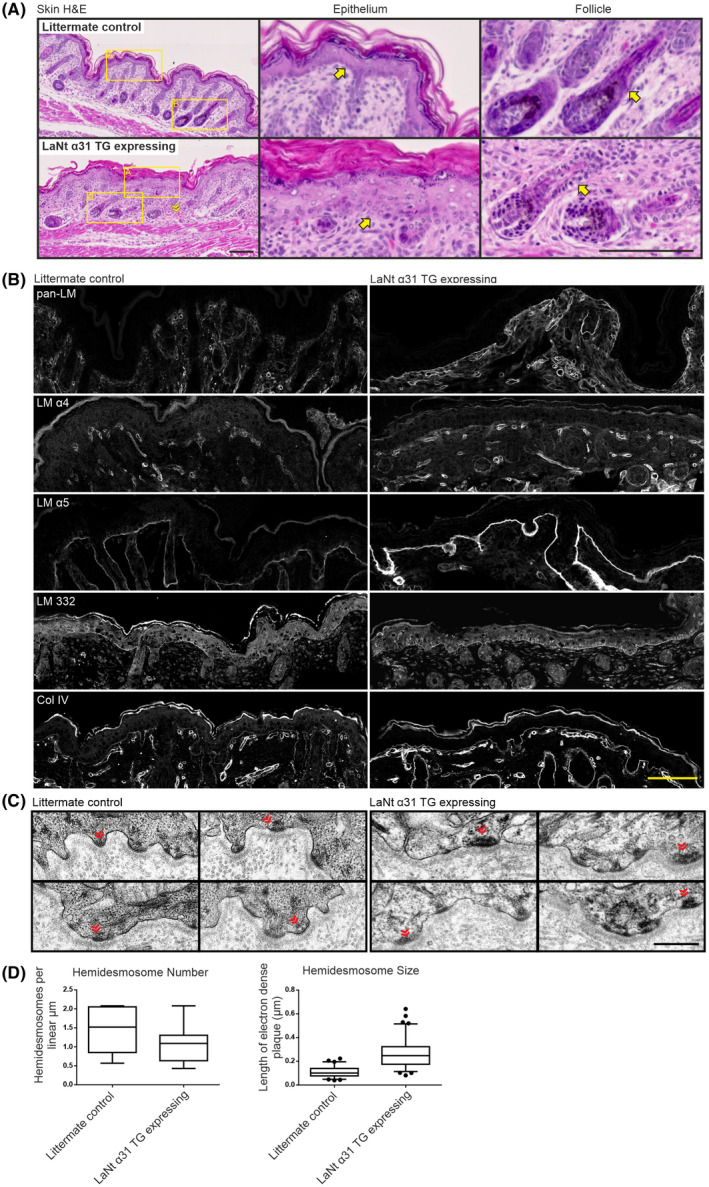
Laminins (LMs) are essential components of all basement membranes where they regulate an extensive array of tissue functions. Alternative splicing from the laminin α3 gene produces a non-laminin but netrin-like protein, Laminin N terminus α31 (LaNt α31). LaNt α31 is widely expressed in intact tissue and is upregulated in epithelial cancers and during wound healing. In vitro functional studies have shown that LaNt α31 can influence numerous aspects of epithelial cell behavior via modifying matrix organization, suggesting a new model of laminin auto-regulation. However, the function of this protein has not been established in vivo. Here, a mouse transgenic line was generated using the ubiquitin C promoter to drive inducible expression of LaNt α31. When expression was induced at embryonic day 15.5, LaNt α31 transgenic animals were not viable at birth, exhibiting localized regions of erythema. Histologically, the most striking defect was widespread evidence of extravascular bleeding across multiple tissues. Additionally, LaNt α31 transgene expressing animals exhibited kidney epithelial detachment, tubular dilation, disruption of the epidermal basal cell layer and of the hair follicle outer root sheath, and ~50% reduction of cell numbers in the liver, associated with depletion of hematopoietic erythrocytic foci. These findings provide the first in vivo evidence that LaNt α31 can influence tissue morphogenesis.
层粘连蛋白(LMs)是所有基底膜的基本组成部分,在这些基底膜中,它们调节着广泛的组织功能。从层粘连蛋白α3 基因的选择性剪接产生一种非层粘连蛋白但类似神经导向因子的蛋白质,即层粘连蛋白 N 端α31(LaNtα31)。LaNtα31 在完整组织中广泛表达,并在上皮癌和伤口愈合过程中上调。体外功能研究表明,LaNtα31 可以通过改变基质组织,影响上皮细胞行为的众多方面,这表明了一种新的层粘连蛋白自身调节模型。然而,这种蛋白质的功能尚未在体内得到证实。在这里,我们使用泛素 C 启动子生成了一种小鼠转基因系,以诱导表达 LaNtα31。当在胚胎第 15.5 天诱导表达时,LaNtα31 转基因动物在出生时无法存活,表现出局部红斑区域。组织学上,最显著的缺陷是广泛存在的血管外出血证据,涉及多个组织。此外,表达 LaNtα31 转基因的动物表现出肾脏上皮细胞脱落、肾小管扩张、表皮基底细胞层和毛囊外鞘破坏,以及肝脏细胞数量减少约 50%,与造血红细胞灶耗竭有关。这些发现提供了首个体内证据,表明 LaNtα31 可以影响组织形态发生。