• 文献检索
  • 文档翻译
  • 深度研究
  • 学术资讯
  • Suppr Zotero 插件Zotero 插件
  • 邀请有礼
  • 套餐&价格
  • 历史记录
应用&插件
Suppr Zotero 插件Zotero 插件浏览器插件Mac 客户端Windows 客户端微信小程序
定价
高级版会员购买积分包购买API积分包
服务
文献检索文档翻译深度研究API 文档MCP 服务
关于我们
关于 Suppr公司介绍联系我们用户协议隐私条款
关注我们

Suppr 超能文献

核心技术专利:CN118964589B侵权必究
粤ICP备2023148730 号-1Suppr @ 2026

文献检索

告别复杂PubMed语法,用中文像聊天一样搜索,搜遍4000万医学文献。AI智能推荐,让科研检索更轻松。

立即免费搜索

文件翻译

保留排版,准确专业,支持PDF/Word/PPT等文件格式,支持 12+语言互译。

免费翻译文档

深度研究

AI帮你快速写综述,25分钟生成高质量综述,智能提取关键信息,辅助科研写作。

立即免费体验

利用高分辨率光相干断层扫描进行黄斑血管成像和连通性分析。

Macular Vascular Imaging and Connectivity Analysis Using High-Resolution Optical Coherence Tomography.

机构信息

Vitreous Retina Macula Consultants of New York, NY, USA.

CEDOC, NOVA Medical School I Faculdade de Ciências Médicas, Universidade NOVA de Lisboa, Lisbon, Portugal.

出版信息

Transl Vis Sci Technol. 2022 Jun 1;11(6):2. doi: 10.1167/tvst.11.6.2.

DOI:10.1167/tvst.11.6.2
PMID:35648637
原文链接:https://pmc.ncbi.nlm.nih.gov/articles/PMC9172017/
Abstract

PURPOSE

To characterize macular blood flow connectivity in vivo using high-resolution optical coherence tomography (HighRes OCT).

METHODS

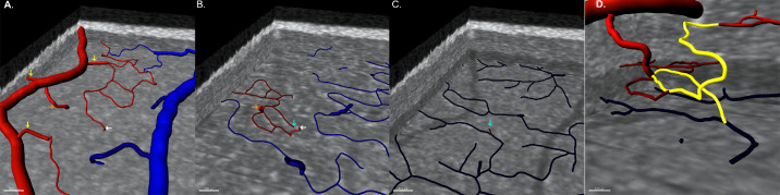
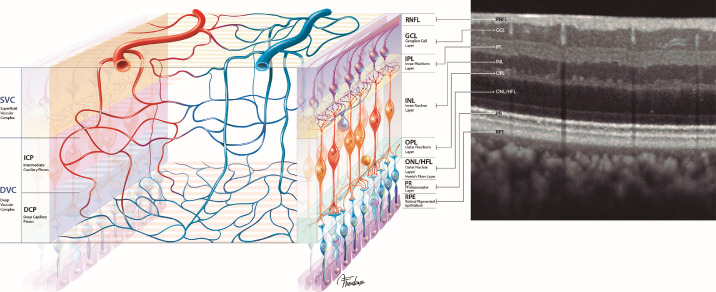
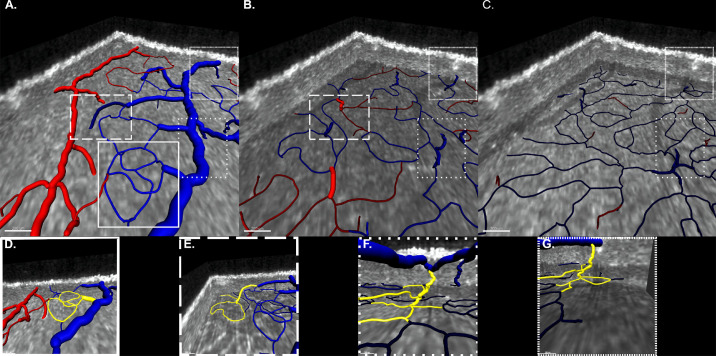
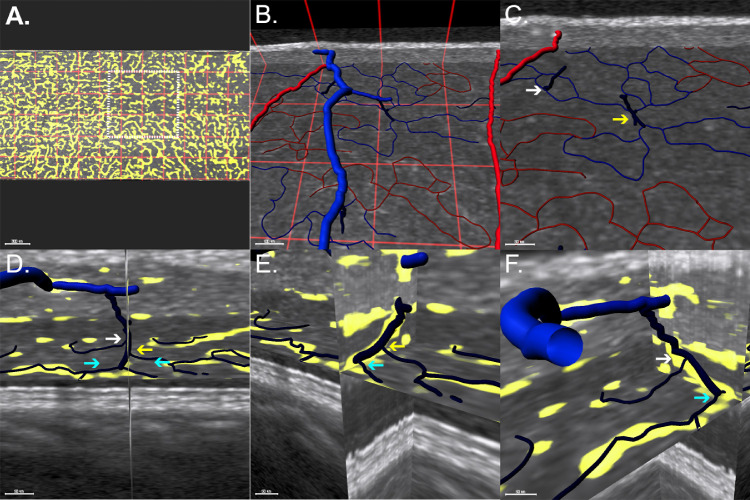
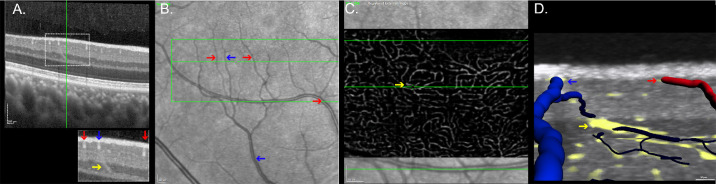
Cross-sectional, observational study. Dense (6-µm interscan distance) perifoveal HighRes OCT raster scans were performed on healthy participants. To mitigate the limitations of projection-resolved OCT-angiography, flow and structural data were used to observe the vascular structures of the superficial vascular complex (SVC) and the deep vascular complex. Vascular segmentation and rendering were performed using Imaris 9.5 software. Inflow and outflow patterns were classified according to vascular diameter and branching order from superficial arteries and veins, respectively.

RESULTS

Eight eyes from eight participants were included in this analysis, from which 422 inflow and 459 outflow connections were characterized. Arteries had direct arteriolar connections to the SVC (78%) and to the intermediate capillary plexus (ICP, 22%). Deep capillary plexus (DCP) inflow derived from small-diameter vessels succeeding ICP arterioles. The most prevalent outflow pathways coursed through superficial draining venules (74%). DCP draining venules ordinarily merged with ICP draining venules and drained independently of superficial venules in 21% of cases. The morphology of DCP draining venules in structural HighRes OCT is distinct from other vessels crossing the inner nuclear layer and can be used to identify superficial veins.

CONCLUSIONS

Vascular connectivity analysis supports a hybrid circuitry of blood flow within the human parafoveal macula.

TRANSLATIONAL RELEVANCE

Characterization of parafoveal macular blood flow connectivity in vivo using a precise segmentation of HighRes OCT is consistent with ground-truth microscopy studies and shows a hybrid circuitry.

摘要

目的

使用高分辨率光学相干断层扫描(HighRes OCT)对活体黄斑血流连接进行特征描述。

方法

这是一项横断面观察性研究。在健康参与者中进行密集(6μm 扫描间隔)的黄斑区 HighRes OCT 光栅扫描。为了减轻投影分辨 OCT 血管造影的局限性,使用血流和结构数据观察浅层血管复合体(SVC)和深层血管复合体的血管结构。使用 Imaris 9.5 软件进行血管分割和渲染。根据从浅层动脉和静脉分支的血管直径和分支顺序,将流入和流出模式进行分类。

结果

本分析纳入了 8 名参与者的 8 只眼,共描述了 422 个流入和 459 个流出连接。动脉与 SVC(78%)和中间毛细血管丛(ICP,22%)的小动脉有直接的小动脉连接。深层毛细血管丛(DCP)的流入来源于 ICP 小动脉之后的小直径血管。最常见的流出途径是通过浅层引流小静脉(74%)。在 21%的情况下,DCP 引流小静脉通常与 ICP 引流小静脉合并,并独立于浅层小静脉引流。结构 HighRes OCT 中 DCP 引流小静脉的形态与穿过内核层的其他血管明显不同,可用于识别浅层静脉。

结论

血管连接分析支持人类旁黄斑区血流的混合电路。

翻译

袁霄霄

https://cdn.ncbi.nlm.nih.gov/pmc/blobs/ce47/9172017/beb8f3c719a1/tvst-11-6-2-f005.jpg
https://cdn.ncbi.nlm.nih.gov/pmc/blobs/ce47/9172017/a954e7659162/tvst-11-6-2-f001.jpg
https://cdn.ncbi.nlm.nih.gov/pmc/blobs/ce47/9172017/a67a11573b27/tvst-11-6-2-f002.jpg
https://cdn.ncbi.nlm.nih.gov/pmc/blobs/ce47/9172017/6cc21e851d04/tvst-11-6-2-f003.jpg
https://cdn.ncbi.nlm.nih.gov/pmc/blobs/ce47/9172017/8ff625b25f3a/tvst-11-6-2-f004.jpg
https://cdn.ncbi.nlm.nih.gov/pmc/blobs/ce47/9172017/beb8f3c719a1/tvst-11-6-2-f005.jpg

相似文献

1
Macular Vascular Imaging and Connectivity Analysis Using High-Resolution Optical Coherence Tomography.利用高分辨率光相干断层扫描进行黄斑血管成像和连通性分析。
Transl Vis Sci Technol. 2022 Jun 1;11(6):2. doi: 10.1167/tvst.11.6.2.
2
Volume Rendering of Dense B-Scan Optical Coherence Tomography Angiography to Evaluate the Connectivity of Macular Blood Flow.基于体绘制的密集 B 扫描光学相干断层血管造影术评估黄斑血流连通性。
Invest Ophthalmol Vis Sci. 2020 Jun 3;61(6):44. doi: 10.1167/iovs.61.6.44.
3
Projection-Resolved Optical Coherence Tomographic Angiography of Retinal Plexuses in Retinitis Pigmentosa.视网膜色素变性的视网膜神经纤维层光学相干断层扫描血管造影术。
Am J Ophthalmol. 2019 Aug;204:70-79. doi: 10.1016/j.ajo.2019.02.034. Epub 2019 Mar 6.
4
OCT Angiography: Measurement of Retinal Macular Microvasculature with Spectralis II OCT Angiography - Reliability and Reproducibility.OCT 血管造影:应用 Spectralis II OCT 血管造影测量视网膜黄斑微循环 - 可靠性和可重复性。
Ophthalmologica. 2020;243(1):75-84. doi: 10.1159/000502458. Epub 2019 Sep 11.
5
Detection of Reduced Retinal Vessel Density in Eyes with Geographic Atrophy Secondary to Age-Related Macular Degeneration Using Projection-Resolved Optical Coherence Tomography Angiography.采用投影分辨率光学相干断层血管造影术检测与年龄相关性黄斑变性相关的地图状萎缩性眼病的视网膜血管密度降低。
Am J Ophthalmol. 2020 Jan;209:206-212. doi: 10.1016/j.ajo.2019.09.004. Epub 2019 Sep 14.
6
Volume Rendering of Deep Retinal Age-Related Microvascular Anomalies.深度视网膜年龄相关性微血管异常的容积再现
Ophthalmol Retina. 2022 Dec;6(12):1185-1193. doi: 10.1016/j.oret.2022.06.005. Epub 2022 Jun 27.
7
OCT-Angiography Comparison between Obstructive Sleep Apnea Children and Normal Subjects in China.中国阻塞性睡眠呼吸暂停儿童与正常儿童的 OCT-Angiography 比较。
Curr Eye Res. 2021 Mar;46(3):355-360. doi: 10.1080/02713683.2020.1801757. Epub 2020 Aug 11.
8
Projection-Resolved Optical Coherence Tomography Angiography of Macular Retinal Circulation in Glaucoma.青光眼黄斑视网膜循环的投影分辨光学相干断层扫描血管造影
Ophthalmology. 2017 Nov;124(11):1589-1599. doi: 10.1016/j.ophtha.2017.06.002. Epub 2017 Jul 1.
9
Assessment of macular microvasculature features before and after vitrectomy in the idiopathic macular epiretinal membrane using a grading system: An optical coherence tomography angiography study.采用分级系统评估特发性黄斑视网膜前膜玻璃体切除术前、后黄斑微血管特征:光学相干断层扫描血管造影研究。
Acta Ophthalmol. 2021 Nov;99(7):e1168-e1175. doi: 10.1111/aos.14753. Epub 2021 Jan 10.
10
Three-Dimensional Characterization of the Normal Human Parafoveal Microvasculature Using Structural Criteria and High-Resolution Confocal Microscopy.利用结构标准和高分辨率共聚焦显微镜对正常人周边视网膜微循环进行三维特征描述。
Invest Ophthalmol Vis Sci. 2020 Aug 3;61(10):3. doi: 10.1167/iovs.61.10.3.

引用本文的文献

1
Paracentral Acute Middle Maculopathy (PAMM) in Ocular Vascular Diseases-What We Know and Future Perspectives.眼部血管疾病中的黄斑中心凹旁急性中层病变(PAMM)——我们所了解的及未来展望
Vision (Basel). 2025 Mar 3;9(1):19. doi: 10.3390/vision9010019.
2
High-Resolution OCT Reveals Age-Associated Variation in the Region Posterior to the External Limiting Membrane.高分辨率光学相干断层扫描揭示了外界膜后方区域与年龄相关的变化。
Transl Vis Sci Technol. 2025 Jan 2;14(1):16. doi: 10.1167/tvst.14.1.16.
3
QUANTIFICATION OF CAPILLARY BLOOD FLOW SPEEDS IN DIABETIC RETINOPATHY USING VARIABLE INTERSCAN TIME ANALYSIS OPTICAL COHERENCE TOMOGRAPHY ANGIOGRAPHY.

本文引用的文献

1
Coincident PAMM and AMN and Insights Into a Common Pathophysiology.同时发生的 PAMM 和 AMN 及对共同病理生理学的深入了解。
Am J Ophthalmol. 2022 Apr;236:136-146. doi: 10.1016/j.ajo.2021.07.004. Epub 2021 Jul 18.
2
INTERMEDIATE AND DEEP CAPILLARY PLEXUSES IN MACHINE LEARNING SEGMENTATION OF HIGH-RESOLUTION OPTICAL COHERENCE TOMOGRAPHY IMAGING.机器学习在高分辨率光学相干断层成像中的中间和深层毛细血管丛分割。
Retina. 2021 Jun 1;41(6):1314-1317. doi: 10.1097/IAE.0000000000003097.
3
Exudative non-neovascular age-related macular degeneration.
使用可变扫描时间分析光学相干断层扫描血管造影术对糖尿病视网膜病变患者的毛细血管血流速度进行定量分析
Retina. 2025 Jan 1;45(1):35-43. doi: 10.1097/IAE.0000000000004279.
4
Acute macular neuroretinopathy following COVID-19 infection: Three case reports.新型冠状病毒肺炎感染后急性黄斑神经视网膜病变:三例报告
World J Clin Cases. 2024 Sep 6;12(25):5775-5783. doi: 10.12998/wjcc.v12.i25.5775.
5
Topographic Measurement of the Subretinal Pigment Epithelium Space in Normal Aging and Age-Related Macular Degeneration Using High-Resolution OCT.使用高分辨率 OCT 对正常衰老和年龄相关性黄斑变性的视网膜色素上皮下空间进行地形测量。
Invest Ophthalmol Vis Sci. 2024 Aug 1;65(10):18. doi: 10.1167/iovs.65.10.18.
6
Optical Coherence Tomography and Optical Coherence Tomography Angiography: Essential Tools for Detecting Glaucoma and Disease Progression.光学相干断层扫描和光学相干断层扫描血管造影:检测青光眼和疾病进展的重要工具。
Front Ophthalmol (Lausanne). 2023;3. doi: 10.3389/fopht.2023.1217125. Epub 2023 Jul 28.
7
Mapping the human parafoveal vascular network to understand flow variability in capillaries.绘制人周边视网膜血管网络图以了解毛细血管中的血流变异性。
PLoS One. 2023 Oct 13;18(10):e0292962. doi: 10.1371/journal.pone.0292962. eCollection 2023.
8
High-Resolution Optical Coherence Tomography in Healthy Individuals Provides Resolution at the Cellular and Subcellular Levels.高分辨率光学相干断层扫描在健康个体中提供细胞和亚细胞水平的分辨率。
Transl Vis Sci Technol. 2023 Jul 3;12(7):12. doi: 10.1167/tvst.12.7.12.
9
Optical Coherence Tomography Angiography of Volumetric Arteriovenous Relationships in the Healthy Macula and Their Derangement in Disease.健康黄斑区容积动静脉关系的光相干断层扫描血管造影及其在疾病中的变化。
Invest Ophthalmol Vis Sci. 2023 May 1;64(5):6. doi: 10.1167/iovs.64.5.6.
10
Histology of Type 3 Macular Neovascularization and Microvascular Anomalies in Treated Age-Related Macular Degeneration: A Case Study.治疗后的年龄相关性黄斑变性中3型黄斑新生血管形成和微血管异常的组织学:一项病例研究
Ophthalmol Sci. 2023 Feb 10;3(3):100280. doi: 10.1016/j.xops.2023.100280. eCollection 2023 Sep.
渗出型非新生血管性年龄相关性黄斑变性。
Graefes Arch Clin Exp Ophthalmol. 2021 May;259(5):1123-1134. doi: 10.1007/s00417-020-05021-y. Epub 2020 Nov 26.
4
Collaborative Filtering of Correlated Noise: Exact Transform-Domain Variance for Improved Shrinkage and Patch Matching.相关噪声的协同过滤:用于改进收缩和补丁匹配的精确变换域方差
IEEE Trans Image Process. 2020 Aug 12;PP. doi: 10.1109/TIP.2020.3014721.
5
Three-Dimensional Characterization of the Normal Human Parafoveal Microvasculature Using Structural Criteria and High-Resolution Confocal Microscopy.利用结构标准和高分辨率共聚焦显微镜对正常人周边视网膜微循环进行三维特征描述。
Invest Ophthalmol Vis Sci. 2020 Aug 3;61(10):3. doi: 10.1167/iovs.61.10.3.
6
Volume Rendering of Dense B-Scan Optical Coherence Tomography Angiography to Evaluate the Connectivity of Macular Blood Flow.基于体绘制的密集 B 扫描光学相干断层血管造影术评估黄斑血流连通性。
Invest Ophthalmol Vis Sci. 2020 Jun 3;61(6):44. doi: 10.1167/iovs.61.6.44.
7
Differentiating Veins From Arteries on Optical Coherence Tomography Angiography by Identifying Deep Capillary Plexus Vortices.通过识别深层毛细血管丛涡旋区分光学相干断层血管造影中的静脉和动脉。
Am J Ophthalmol. 2019 Nov;207:363-372. doi: 10.1016/j.ajo.2019.06.009. Epub 2019 Jun 19.
8
Hemodynamic Response of the Three Macular Capillary Plexuses in Dark Adaptation and Flicker Stimulation Using Optical Coherence Tomography Angiography.利用光相干断层扫描血管造影术观察暗适应和闪烁刺激下三个黄斑毛细血管丛的血流动力学反应。
Invest Ophthalmol Vis Sci. 2019 Feb 1;60(2):694-703. doi: 10.1167/iovs.18-25478.
9
Rodent retinal circulation organization and oxygen metabolism revealed by visible-light optical coherence tomography.可见光光学相干断层扫描揭示的啮齿动物视网膜循环组织与氧代谢
Biomed Opt Express. 2018 Oct 30;9(11):5851-5862. doi: 10.1364/BOE.9.005851. eCollection 2018 Nov 1.
10
Association of Optical Coherence Tomography Angiography of Collaterals in Retinal Vein Occlusion With Major Venous Outflow Through the Deep Vascular Complex.视网膜静脉阻塞患者侧支循环的光学相干断层血管造影与深血管复合体主要静脉流出的相关性。
JAMA Ophthalmol. 2018 Nov 1;136(11):1262-1270. doi: 10.1001/jamaophthalmol.2018.3586.