Berlin Andreas, Cabral Diogo, Chen Ling, Messinger Jeffrey D, Balaratnasingam Chandrakumar, Mendis Randev, Ferrara Daniela, Freund K Bailey, Curcio Christine A
Department of Ophthalmology and Visual Sciences, Heersink School of Medicine, University of Alabama at Birmingham, Birmingham, Alabama.
Department of Ophthalmology, University Hospital Wurzburg, Wurzburg, Germany.
Ophthalmol Sci. 2023 Feb 10;3(3):100280. doi: 10.1016/j.xops.2023.100280. eCollection 2023 Sep.
To investigate intraretinal neovascularization and microvascular anomalies by correlating in vivo multimodal imaging with corresponding ex vivo histology in a single patient.
A case study comprising clinical imaging from a community-based practice, and histologic analysis at a university-based research laboratory (clinicopathologic correlation).
A White woman in her 90s treated with numerous intravitreal anti-VEGF injections for bilateral type 3 macular neovascularization (MNV) secondary to age-related macular degeneration (AMD).
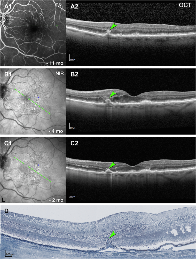
Clinical imaging comprised serial infrared reflectance, eye-tracked spectral-domain OCT, OCT angiography, and fluorescein angiography. Eye tracking, applied to the 2 preserved donor eyes, enabled the correlation of clinical imaging signatures with high-resolution histology and transmission electron microscopy.
Histologic/ultrastructural descriptions and diameters of vessels seen in clinical imaging.
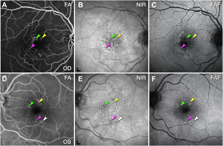
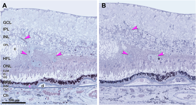
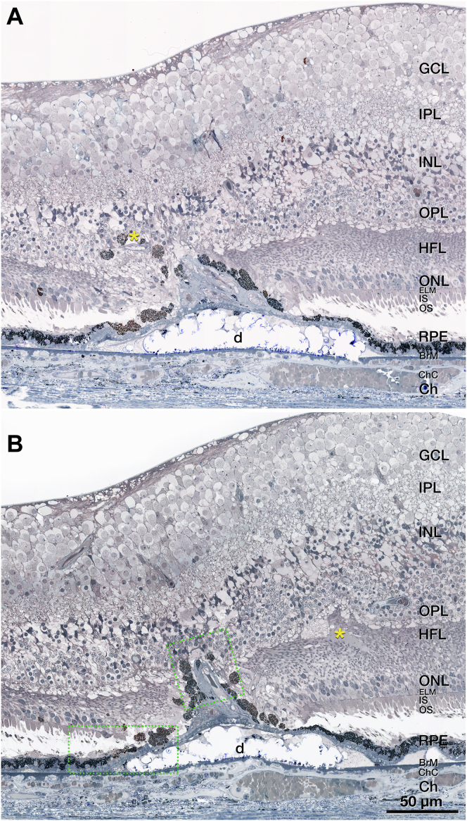
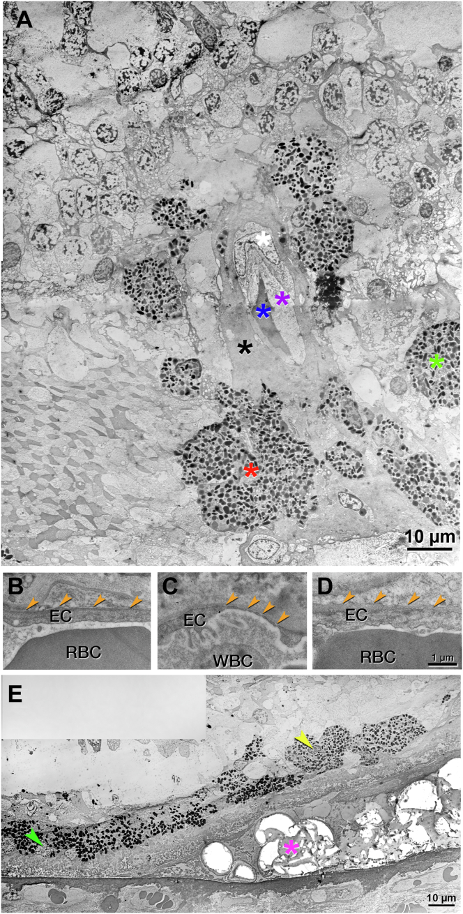
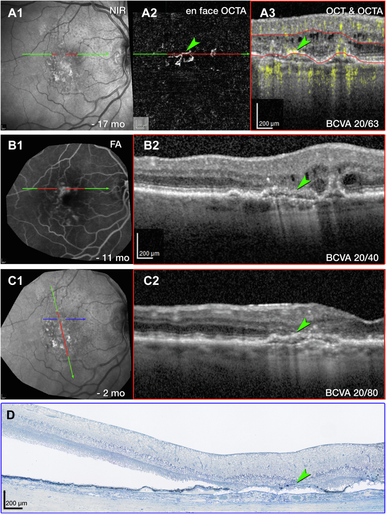
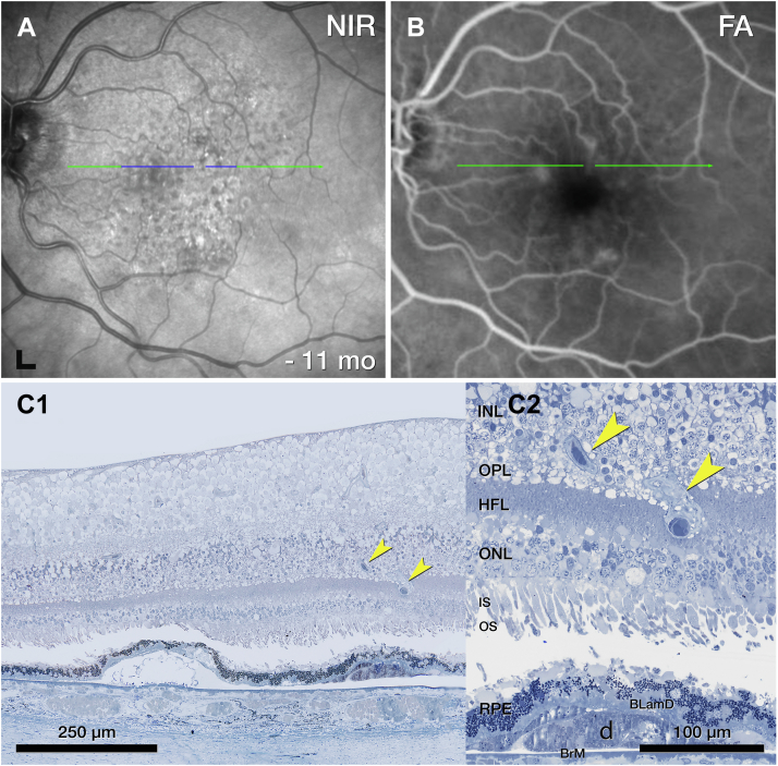
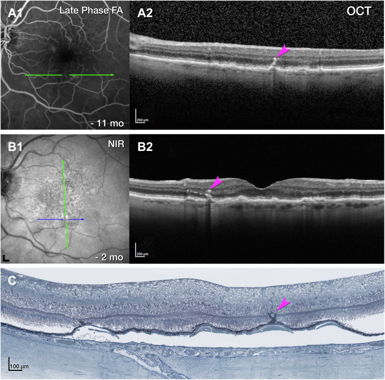
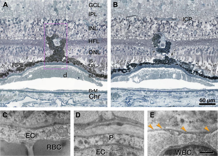
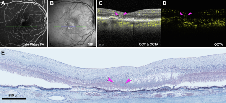
Six vascular lesions were histologically confirmed (type 3 MNV, n = 3; deep retinal age-related microvascular anomalies [DRAMAs], n = 3). Pyramidal (n = 2) or tangled (n = 1) morphologies of type 3 MNV originated at the deep capillary plexus (DCP) and extended posteriorly to approach without penetrating persistent basal laminar deposit. They did not enter the subretinal pigment epithelium (RPE)-basal laminar space or cross the Bruch membrane. Choroidal contributions were not found. The neovascular complexes included pericytes and nonfenestrated endothelial cells, within a collagenous sheath covered by dysmorphic RPE cells. Deep retinal age-related microvascular anomaly lesions extended posteriorly from the DCP into the Henle fiber and the outer nuclear layers without evidence of atrophy, exudation, or anti-VEGF responsiveness. Two DRAMAs lacked collagenous sheaths. External and internal diameters of type 3 MNV and DRAMA vessels were larger than comparison vessels in the index eyes and in aged normal and intermediate AMD eyes.
Type 3 MNV vessels reflect specializations of source capillaries and persist during anti-VEGF therapy. The collagenous sheath of type 3 MNV lesions may provide structural stabilization. If so, vascular characteristics may be useful in disease monitoring in addition to fluid and flow signal detection. Further investigation with longitudinal imaging before exudation onset will help determine if DRAMAs are part of the type 3 MNV progression sequence.
Proprietary or commercial disclosure may be found after the references.
通过将一名患者的体内多模态成像与相应的体外组织学进行关联,研究视网膜内新生血管形成和微血管异常情况。
一项病例研究,包括来自社区诊所的临床成像以及大学研究实验室的组织学分析(临床病理相关性研究)。
一名90多岁的白人女性,因年龄相关性黄斑变性(AMD)继发双侧3型黄斑新生血管(MNV)接受了多次玻璃体内抗VEGF注射治疗。
临床成像包括连续红外反射、眼跟踪光谱域光学相干断层扫描(OCT)、OCT血管造影和荧光素血管造影。对2只保存的供体眼应用眼跟踪技术,使临床成像特征与高分辨率组织学和透射电子显微镜相关联。
临床成像中所见血管的组织学/超微结构描述及直径。
组织学证实有6个血管病变(3型MNV,n = 3;深部视网膜年龄相关性微血管异常[DRAMAs],n = 3)。3型MNV的金字塔形(n = 2)或缠结形(n = 1)形态起源于深部毛细血管丛(DCP),向后延伸接近但未穿透持续的基底膜沉积物。它们未进入视网膜色素上皮(RPE)-基底膜间隙或穿过布鲁赫膜。未发现脉络膜的参与。新生血管复合体包括周细胞和无窗孔内皮细胞,位于由形态异常的RPE细胞覆盖的胶原鞘内。深部视网膜年龄相关性微血管异常病变从DCP向后延伸至Henle纤维层和外核层,无萎缩、渗出或抗VEGF反应的证据。2个DRAMAs缺乏胶原鞘。3型MNV和DRAMA血管的外径和内径均大于患眼以及年龄匹配的正常眼和中度AMD眼中的对照血管。
3型MNV血管反映了源毛细血管的特殊化,并在抗VEGF治疗期间持续存在。3型MNV病变的胶原鞘可能提供结构稳定性。如果是这样,除了检测液体和血流信号外,血管特征可能有助于疾病监测。在渗出发作前进行纵向成像的进一步研究将有助于确定DRAMAs是否是3型MNV进展序列的一部分。
在参考文献之后可能会有专利或商业披露信息。