• 文献检索
  • 文档翻译
  • 深度研究
  • 学术资讯
  • Suppr Zotero 插件Zotero 插件
  • 邀请有礼
  • 套餐&价格
  • 历史记录
应用&插件
Suppr Zotero 插件Zotero 插件浏览器插件Mac 客户端Windows 客户端微信小程序
定价
高级版会员购买积分包购买API积分包
服务
文献检索文档翻译深度研究API 文档MCP 服务
关于我们
关于 Suppr公司介绍联系我们用户协议隐私条款
关注我们

Suppr 超能文献

核心技术专利:CN118964589B侵权必究
粤ICP备2023148730 号-1Suppr @ 2026

文献检索

告别复杂PubMed语法,用中文像聊天一样搜索,搜遍4000万医学文献。AI智能推荐,让科研检索更轻松。

立即免费搜索

文件翻译

保留排版,准确专业,支持PDF/Word/PPT等文件格式,支持 12+语言互译。

免费翻译文档

深度研究

AI帮你快速写综述,25分钟生成高质量综述,智能提取关键信息,辅助科研写作。

立即免费体验

多 sclerosis 中感觉运动皮层病灶与对侧手功能的关联:7 T MRI 研究。

Linking lesions in sensorimotor cortex to contralateral hand function in multiple sclerosis: a 7 T MRI study.

机构信息

Danish Research Centre for Magnetic Resonance, Copenhagen University Hospital-Amager and Hvidovre, 2650 Hvidovre, Denmark.

Department of Health Technology, Technical University of Denmark, 2800 Kgs Lyngby, Denmark.

出版信息

Brain. 2022 Oct 21;145(10):3522-3535. doi: 10.1093/brain/awac203.

DOI:10.1093/brain/awac203
PMID:35653498
原文链接:https://pmc.ncbi.nlm.nih.gov/articles/PMC9586550/
Abstract

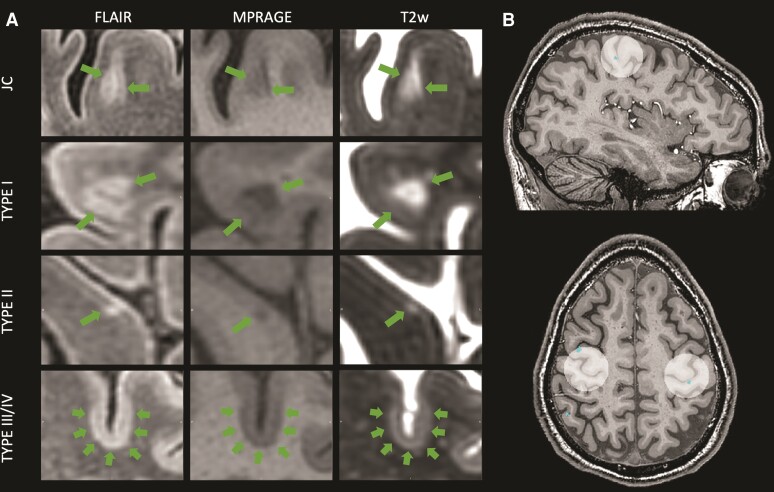
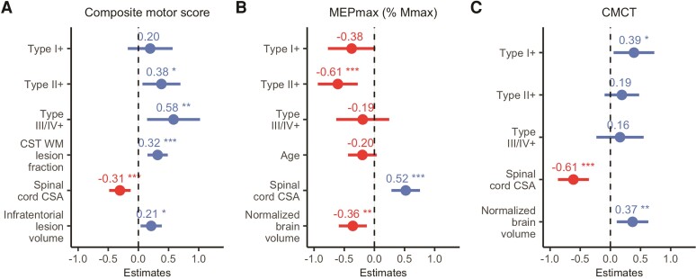
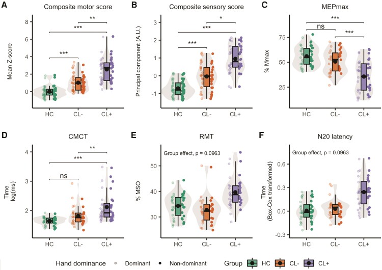
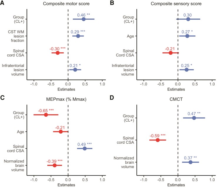
Cortical lesions constitute a key manifestation of multiple sclerosis and contribute to clinical disability and cognitive impairment. Yet it is unknown whether local cortical lesions and cortical lesion subtypes contribute to domain-specific impairments attributable to the function of the lesioned cortex. In this cross-sectional study, we assessed how cortical lesions in the primary sensorimotor hand area relate to corticomotor physiology and sensorimotor function of the contralateral hand. Fifty relapse-free patients with relapsing-remitting or secondary-progressive multiple sclerosis and 28 healthy age- and sex-matched participants underwent whole-brain 7 T MRI to map cortical lesions. Brain scans were also used to estimate normalized brain volume, pericentral cortical thickness, white matter lesion fraction of the corticospinal tract, infratentorial lesion volume and the cross-sectional area of the upper cervical spinal cord. We tested sensorimotor hand function and calculated a motor and sensory composite score for each hand. In 37 patients and 20 healthy controls, we measured maximal motor-evoked potential amplitude, resting motor threshold and corticomotor conduction time with transcranial magnetic stimulation and the N20 latency from somatosensory-evoked potentials. Patients showed at least one cortical lesion in the primary sensorimotor hand area in 47 of 100 hemispheres. The presence of a lesion was associated with worse contralateral sensory (P = 0.014) and motor (P = 0.009) composite scores. Transcranial magnetic stimulation of a lesion-positive primary sensorimotor hand area revealed a decreased maximal motor-evoked potential amplitude (P < 0.001) and delayed corticomotor conduction (P = 0.002) relative to a lesion-negative primary sensorimotor hand area. Stepwise mixed linear regressions showed that the presence of a primary sensorimotor hand area lesion, higher white-matter lesion fraction of the corticospinal tract, reduced spinal cord cross-sectional area and higher infratentorial lesion volume were associated with reduced contralateral motor hand function. Cortical lesions in the primary sensorimotor hand area, spinal cord cross-sectional area and normalized brain volume were also associated with smaller maximal motor-evoked potential amplitude and longer corticomotor conduction times. The effect of cortical lesions on sensory function was no longer significant when controlling for MRI-based covariates. Lastly, we found that intracortical and subpial lesions had the largest effect on reduced motor hand function, intracortical lesions on reduced motor-evoked potential amplitude and leucocortical lesions on delayed corticomotor conduction. Together, this comprehensive multilevel assessment of sensorimotor brain damage shows that the presence of a cortical lesion in the primary sensorimotor hand area is associated with impaired corticomotor function of the hand, after accounting for damage at the subcortical level. The results also provide preliminary evidence that cortical lesion types may affect the various facets of corticomotor function differentially.

摘要

皮质病变是多发性硬化症的一个重要表现,并导致临床残疾和认知障碍。然而,目前尚不清楚局部皮质病变和皮质病变亚型是否会导致与病变皮质功能相关的特定领域的损伤。在这项横断面研究中,我们评估了初级感觉运动手区的皮质病变与对侧手的皮质运动生理学和感觉运动功能之间的关系。50 名无复发缓解或继发进展性多发性硬化症患者和 28 名年龄和性别匹配的健康参与者接受了全脑 7T MRI 以绘制皮质病变图。脑扫描还用于估计标准化脑体积、皮质下中央区皮质厚度、皮质脊髓束的白质病变分数、颅后窝病变体积和上颈段脊髓的横截面积。我们测试了手的感觉运动功能,并为每只手计算了运动和感觉综合评分。在 37 名患者和 20 名健康对照者中,我们使用经颅磁刺激测量了最大运动诱发电位幅度、静息运动阈值和皮质运动传导时间,以及体感诱发电位的 N20 潜伏期。在 100 个半脑中,有 47 个存在至少一个初级感觉运动手区的皮质病变。病变的存在与对侧感觉(P=0.014)和运动(P=0.009)综合评分较差有关。对皮质病变阳性的初级感觉运动手区进行经颅磁刺激显示,与皮质病变阴性的初级感觉运动手区相比,最大运动诱发电位幅度降低(P<0.001),皮质运动传导时间延迟(P=0.002)。逐步混合线性回归显示,初级感觉运动手区皮质病变的存在、皮质脊髓束的白质病变分数较高、脊髓横截面积减小和颅后窝病变体积增大与对侧手部运动功能下降有关。皮质病变、脊髓横截面积和标准化脑体积也与最大运动诱发电位幅度减小和皮质运动传导时间延长有关。当控制基于 MRI 的协变量时,皮质病变对感觉功能的影响不再显著。最后,我们发现皮质内和皮质下病变对手部运动功能的影响最大,皮质内病变对运动诱发电位幅度的影响最大,白质病变对皮质运动传导的影响最大。综上所述,这种对手运动脑损伤的全面多层次评估表明,在手的皮质运动功能受损时,初级感觉运动手区的皮质病变与病变有关,在考虑到皮质下损伤后。结果还初步表明,皮质病变类型可能会以不同的方式影响皮质运动功能的各个方面。

https://cdn.ncbi.nlm.nih.gov/pmc/blobs/0abe/9586550/87e6166bd0e4/awac203f4.jpg
https://cdn.ncbi.nlm.nih.gov/pmc/blobs/0abe/9586550/f95fa7a141e2/awac203f1.jpg
https://cdn.ncbi.nlm.nih.gov/pmc/blobs/0abe/9586550/6767f9cec791/awac203f2.jpg
https://cdn.ncbi.nlm.nih.gov/pmc/blobs/0abe/9586550/d214b1e3256d/awac203f3.jpg
https://cdn.ncbi.nlm.nih.gov/pmc/blobs/0abe/9586550/87e6166bd0e4/awac203f4.jpg

相似文献

1
Linking lesions in sensorimotor cortex to contralateral hand function in multiple sclerosis: a 7 T MRI study.多 sclerosis 中感觉运动皮层病灶与对侧手功能的关联:7 T MRI 研究。
Brain. 2022 Oct 21;145(10):3522-3535. doi: 10.1093/brain/awac203.
2
Multiple sclerosis lesions in motor tracts from brain to cervical cord: spatial distribution and correlation with disability.脑至颈髓运动通路上多发性硬化病灶:空间分布与残疾的相关性。
Brain. 2020 Jul 1;143(7):2089-2105. doi: 10.1093/brain/awaa162.
3
MRI investigation of the sensorimotor cortex and the corticospinal tract after acute spinal cord injury: a prospective longitudinal study.急性脊髓损伤后感觉运动皮层和皮质脊髓束的 MRI 研究:一项前瞻性纵向研究。
Lancet Neurol. 2013 Sep;12(9):873-881. doi: 10.1016/S1474-4422(13)70146-7. Epub 2013 Jul 2.
4
7 T imaging reveals a gradient in spinal cord lesion distribution in multiple sclerosis.7T 成像显示多发性硬化症的脊髓病变分布呈梯度变化。
Brain. 2020 Oct 1;143(10):2973-2987. doi: 10.1093/brain/awaa249.
5
Differences in brain structure and theta burst stimulation-induced plasticity implicate the corticomotor system in loss of function after musculoskeletal injury.骨骼肌肉损伤后功能丧失与皮质运动系统有关,其与大脑结构和 theta 爆发刺激诱导的可塑性存在差异。
J Neurophysiol. 2021 Apr 1;125(4):1006-1021. doi: 10.1152/jn.00689.2020. Epub 2021 Feb 17.
6
Motor evoked potentials and disability in secondary progressive multiple sclerosis.继发进展型多发性硬化症中的运动诱发电位与残疾情况
Can J Neurol Sci. 1997 Nov;24(4):332-7. doi: 10.1017/s0317167100033011.
7
Association of Cortical Lesion Burden on 7-T Magnetic Resonance Imaging With Cognition and Disability in Multiple Sclerosis.7-T磁共振成像上皮质病变负荷与多发性硬化症认知及残疾的相关性
JAMA Neurol. 2015 Sep;72(9):1004-12. doi: 10.1001/jamaneurol.2015.1241.
8
Sensorimotor dysfunction in multiple sclerosis and column-specific magnetization transfer-imaging abnormalities in the spinal cord.多发性硬化中的感觉运动功能障碍与脊髓中特定柱的磁化传递成像异常。
Brain. 2009 May;132(Pt 5):1200-9. doi: 10.1093/brain/awp032. Epub 2009 Mar 18.
9
Joint assessment of brain and spinal cord motor tract damage in patients with early RRMS: predominant impact of spinal cord lesions on motor function.早期 RRMS 患者脑和脊髓运动束损伤的联合评估:脊髓病变对运动功能的主要影响。
J Neurol. 2019 Sep;266(9):2294-2303. doi: 10.1007/s00415-019-09419-5. Epub 2019 Jun 7.
10
Intracortical inhibition abnormality during the remission phase of multiple sclerosis is related to upper limb dexterity and lesions.多发性硬化缓解期的皮质内抑制异常与上肢灵活性及病灶有关。
Clin Neurophysiol. 2016 Feb;127(2):1503-1511. doi: 10.1016/j.clinph.2015.08.011. Epub 2015 Aug 31.

引用本文的文献

1
The corticospinal tract in multiple sclerosis: correlation between cortical excitability and magnetic resonance imaging measures.多发性硬化症中的皮质脊髓束:皮质兴奋性与磁共振成像测量之间的相关性。
J Neural Transm (Vienna). 2025 Feb;132(2):265-273. doi: 10.1007/s00702-024-02849-0. Epub 2024 Oct 17.
2
Association of Cortical Lesions With Regional Glutamate, GABA, -Acetylaspartate, and Myoinositol Levels in Patients With Multiple Sclerosis.多发性硬化症患者皮质病变与局部谷氨酸、γ-氨基丁酸、N-乙酰天门冬氨酸和肌醇水平的关联
Neurology. 2024 Jul;103(1):e209543. doi: 10.1212/WNL.0000000000209543. Epub 2024 Jun 13.
3
The importance of pyramidal tract integrity for cortical plasticity and related functionality in patients with multiple sclerosis.

本文引用的文献

1
Cortical lesion hotspots and association of subpial lesions with disability in multiple sclerosis.皮质病变热点与多发性硬化症软脑膜下病变相关性及其与残疾的关系。
Mult Scler. 2022 Aug;28(9):1351-1363. doi: 10.1177/13524585211069167. Epub 2022 Feb 10.
2
Imaging cortical multiple sclerosis lesions with ultra-high field MRI.超高场 MRI 成像皮质多发性硬化病变。
Neuroimage Clin. 2021;32:102847. doi: 10.1016/j.nicl.2021.102847. Epub 2021 Oct 6.
3
Acute and chronic synaptic pathology in multiple sclerosis gray matter.多发性硬化症灰质中的急性和慢性突触病理学
锥体束完整性对多发性硬化症患者皮质可塑性及相关功能的重要性。
Front Neurol. 2023 Nov 24;14:1266225. doi: 10.3389/fneur.2023.1266225. eCollection 2023.
4
Long-term three-photon imaging reveals region-specific differences in healthy and regenerative oligodendrogenesis.长期三光子成像揭示了健康和再生少突胶质细胞生成中的区域特异性差异。
bioRxiv. 2023 Nov 1:2023.10.29.564636. doi: 10.1101/2023.10.29.564636.
5
Clinical diagnostic utility of transcranial magnetic stimulation in neurological disorders. Updated report of an IFCN committee.经颅磁刺激在神经障碍中的临床诊断效用。IFCN 委员会的最新报告。
Clin Neurophysiol. 2023 Jun;150:131-175. doi: 10.1016/j.clinph.2023.03.010. Epub 2023 Mar 29.
Mult Scler. 2022 Mar;28(3):369-382. doi: 10.1177/13524585211022174. Epub 2021 Jun 14.
4
A novel prognostic score to assess the risk of progression in relapsing-remitting multiple sclerosis patients.一种用于评估复发缓解型多发性硬化症患者病情进展风险的新型预后评分。
Eur J Neurol. 2021 Aug;28(8):2503-2512. doi: 10.1111/ene.14859. Epub 2021 May 5.
5
CSF parvalbumin levels reflect interneuron loss linked with cortical pathology in multiple sclerosis.脑脊液中 parvalbumin 水平反映了多发性硬化症中与皮质病变相关的中间神经元丢失。
Ann Clin Transl Neurol. 2021 Mar;8(3):534-547. doi: 10.1002/acn3.51298. Epub 2021 Jan 23.
6
Selective vulnerability of inhibitory networks in multiple sclerosis.多发性硬化症中抑制性网络的选择性易损性。
Acta Neuropathol. 2021 Mar;141(3):415-429. doi: 10.1007/s00401-020-02258-z. Epub 2021 Jan 15.
7
A contrast-adaptive method for simultaneous whole-brain and lesion segmentation in multiple sclerosis.一种用于多发性硬化症的全脑和病灶同时分割的对比自适应方法。
Neuroimage. 2021 Jan 15;225:117471. doi: 10.1016/j.neuroimage.2020.117471. Epub 2020 Oct 22.
8
7 T imaging reveals a gradient in spinal cord lesion distribution in multiple sclerosis.7T 成像显示多发性硬化症的脊髓病变分布呈梯度变化。
Brain. 2020 Oct 1;143(10):2973-2987. doi: 10.1093/brain/awaa249.
9
MRI correlates of clinical disability and hand-motor performance in multiple sclerosis phenotypes.MRI 相关性研究:多发性硬化症各表型的临床残疾和手部运动表现
Mult Scler. 2021 Jul;27(8):1205-1221. doi: 10.1177/1352458520958356. Epub 2020 Sep 14.
10
Histopathology-validated recommendations for cortical lesion imaging in multiple sclerosis.组织病理学验证的多发性硬化皮质病变影像学推荐。
Brain. 2020 Oct 1;143(10):2988-2997. doi: 10.1093/brain/awaa233.