• 文献检索
  • 文档翻译
  • 深度研究
  • 学术资讯
  • Suppr Zotero 插件Zotero 插件
  • 邀请有礼
  • 套餐&价格
  • 历史记录
应用&插件
Suppr Zotero 插件Zotero 插件浏览器插件Mac 客户端Windows 客户端微信小程序
定价
高级版会员购买积分包购买API积分包
服务
文献检索文档翻译深度研究API 文档MCP 服务
关于我们
关于 Suppr公司介绍联系我们用户协议隐私条款
关注我们

Suppr 超能文献

核心技术专利:CN118964589B侵权必究
粤ICP备2023148730 号-1Suppr @ 2026

文献检索

告别复杂PubMed语法,用中文像聊天一样搜索,搜遍4000万医学文献。AI智能推荐,让科研检索更轻松。

立即免费搜索

文件翻译

保留排版,准确专业,支持PDF/Word/PPT等文件格式,支持 12+语言互译。

免费翻译文档

深度研究

AI帮你快速写综述,25分钟生成高质量综述,智能提取关键信息,辅助科研写作。

立即免费体验

非侵入性 3T MR 淋巴造影术显示脂肪性脂肪瘤中的皮下脂肪组织水肿。

Subcutaneous Adipose Tissue Edema in Lipedema Revealed by Noninvasive 3T MR Lymphangiography.

机构信息

Department of Radiology and Radiological Sciences, Vanderbilt University Medical Center, Nashville, Tennessee, USA.

Vanderbilt University Institute of Imaging Science, Vanderbilt University Medical Center, Nashville, Tennessee, USA.

出版信息

J Magn Reson Imaging. 2023 Feb;57(2):598-608. doi: 10.1002/jmri.28281. Epub 2022 Jun 3.

DOI:10.1002/jmri.28281
PMID:35657120
原文链接:https://pmc.ncbi.nlm.nih.gov/articles/PMC9718889/
Abstract

BACKGROUND

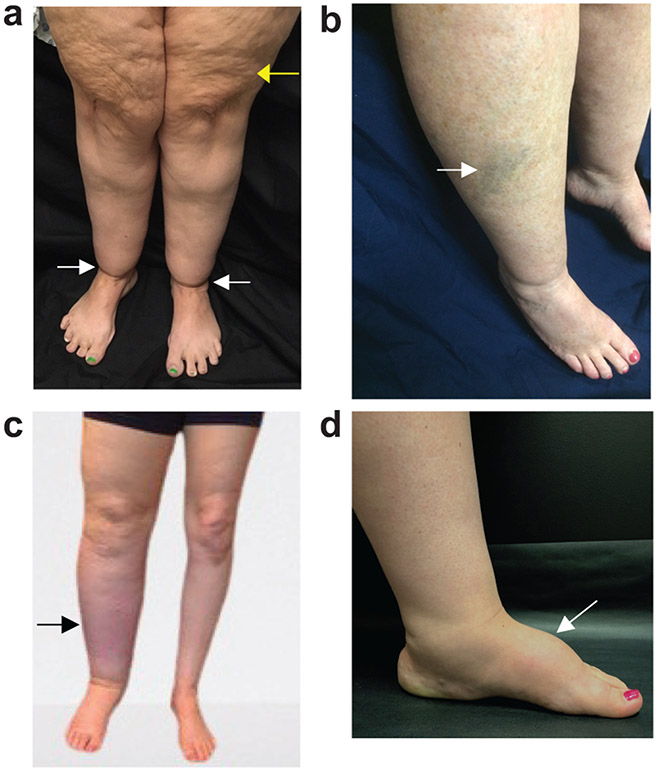
Lipedema exhibits excessive lower-extremity subcutaneous adipose tissue (SAT) deposition, which is frequently misidentified as obesity until lymphedema presents. MR lymphangiography may have relevance to distinguish lipedema from obesity or lymphedema.

HYPOTHESIS

Hyperintensity profiles on 3T MR lymphangiography can identify distinct features consistent with SAT edema in participants with lipedema.

STUDY TYPE

Prospective cross-sectional study.

SUBJECTS

Participants (48 females, matched for age [mean = 44.8 years]) with lipedema (n = 14), lipedema with lymphedema (LWL, n = 12), cancer treatment-related lymphedema (lymphedema, n = 8), and controls without these conditions (n = 14).

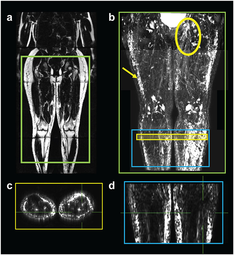
FIELD STRENGTH/SEQUENCE: 3T MR lymphangiography (nontracer 3D turbo-spin-echo).

ASSESSMENT

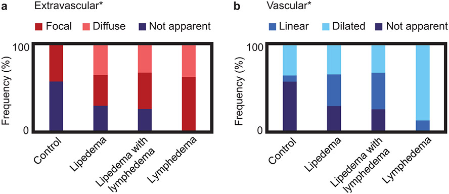
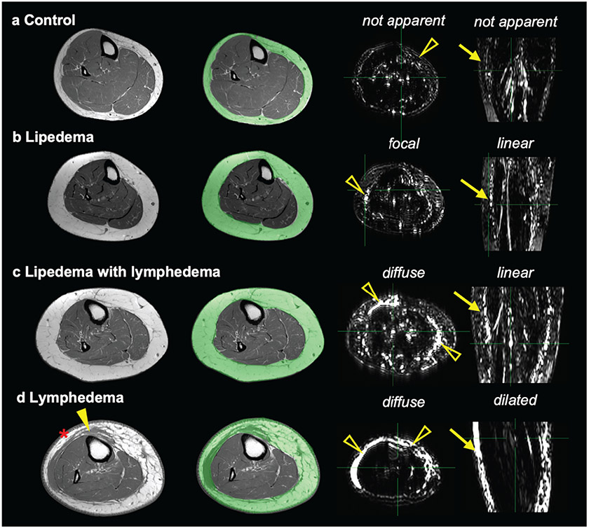
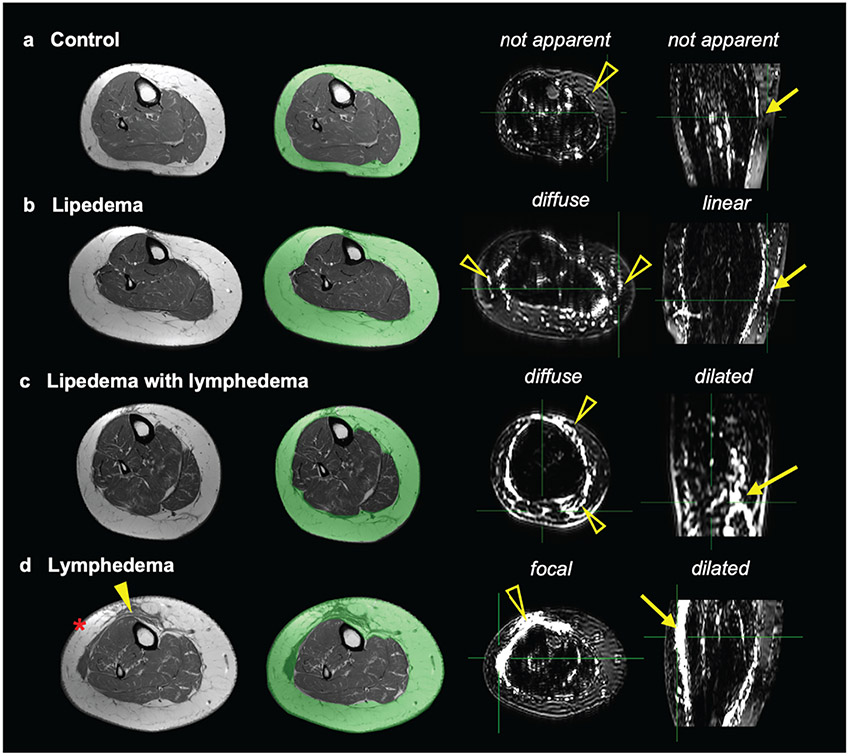
Review of lymphangiograms in lower extremities by three radiologists was performed independently. Spatial patterns of hyperintense signal within the SAT were scored for extravascular (focal, diffuse, or not apparent) and vascular (linear, dilated, or not apparent) image features.

STATISTICAL TESTS

Interreader reliability was computed using Fleiss Kappa. Fisher's exact test was used to evaluate the proportion of image features between study groups. Multinomial logistic regression was used to assess the relationship between image features and study groups. The odds ratio (OR) and 95% confidence interval (CI) of SAT extravascular and vascular features was reported in groups compared to lipedema. The threshold of statistical significance was P < 0.05.

RESULTS

Reliable agreement was demonstrated between three independent, blinded reviewers (P < 0.001). The frequency of SAT hyperintensities in participants with lipedema (36% focal, 36% diffuse), LWL (42% focal, 33% diffuse), lymphedema (62% focal, 38% diffuse), and controls (43% focal, 0% diffuse) was significantly distinct. Compared with lipedema, SAT hyperintensities were less frequent in controls (focal: OR = 0.63, CI = 0.11-3.41; diffuse: OR = 0.05, CI = 0.00-1.27), similar in LWL (focal: OR = 1.29, CI = 0.19-8.89; diffuse: OR = 1.05, CI = 0.15-7.61), and more frequent in lymphedema (focal: OR = 9.00, CI = 0.30-274.12; diffuse: OR = 5.73, CI = 0.18-186.84).

DATA CONCLUSION

Noninvasive MR lymphangiography identifies distinct signal patterns indicating SAT edema and lymphatic load in participants with lipedema.

EVIDENCE LEVEL

1 TECHNICAL EFFICACY: Stage 1.

摘要

背景

脂肪水肿表现为下肢皮下脂肪组织(SAT)过度沉积,直到出现淋巴水肿才常被误认为肥胖。磁共振淋巴管造影术(MR 淋巴造影)可能有助于区分脂肪水肿与肥胖或淋巴水肿。

假说

3T MR 淋巴造影上的高信号特征可识别出与脂肪水肿患者 SAT 水肿一致的特征。

研究类型

前瞻性横断面研究。

受试者

参与者(48 名女性,年龄匹配[平均 44.8 岁]),包括脂肪水肿患者(n=14)、脂肪水肿伴淋巴水肿(LWL,n=12)、癌症治疗相关淋巴水肿(淋巴水肿,n=8)和无这些情况的对照组(n=14)。

磁场强度/序列:3T MR 淋巴造影(非示踪剂 3D 涡轮自旋回波)。

评估

由三位放射科医生独立评估下肢淋巴造影。对 SAT 内高信号的血管外(局灶性、弥漫性或不明显)和血管(线性、扩张或不明显)图像特征进行评分。

统计学检验

采用 Fleiss Kappa 计算读者间可靠性。Fisher 确切检验用于评估研究组之间图像特征的比例。多变量逻辑回归用于评估图像特征与研究组之间的关系。报告了 SAT 血管外和血管特征在各研究组与脂肪水肿患者比较的比值比(OR)和 95%置信区间(CI)。统计显著性阈值为 P<0.05。

结果

三位独立、盲法的评审员之间显示出可靠的一致性(P<0.001)。脂肪水肿患者(36%局灶性,36%弥漫性)、LWL(42%局灶性,33%弥漫性)、淋巴水肿(62%局灶性,38%弥漫性)和对照组(43%局灶性,0%弥漫性)的 SAT 高信号频率存在显著差异。与脂肪水肿相比,对照组 SAT 高信号的频率较低(局灶性:OR=0.63,CI=0.11-3.41;弥漫性:OR=0.05,CI=0.00-1.27),LWL 相似(局灶性:OR=1.29,CI=0.19-8.89;弥漫性:OR=1.05,CI=0.15-7.61),而淋巴水肿的频率较高(局灶性:OR=9.00,CI=0.30-274.12;弥漫性:OR=5.73,CI=0.18-186.84)。

数据结论

非侵入性磁共振淋巴管造影术可识别出指示 SAT 水肿和脂肪水肿患者淋巴负荷的特征性信号模式。

证据水平

1 技术功效:阶段 1。

https://cdn.ncbi.nlm.nih.gov/pmc/blobs/436f/9718889/2859509499ef/nihms-1809599-f0005.jpg
https://cdn.ncbi.nlm.nih.gov/pmc/blobs/436f/9718889/382dff29d6ae/nihms-1809599-f0001.jpg
https://cdn.ncbi.nlm.nih.gov/pmc/blobs/436f/9718889/053efecdfac9/nihms-1809599-f0002.jpg
https://cdn.ncbi.nlm.nih.gov/pmc/blobs/436f/9718889/84a161236dfe/nihms-1809599-f0003.jpg
https://cdn.ncbi.nlm.nih.gov/pmc/blobs/436f/9718889/6c3dbd44819f/nihms-1809599-f0004.jpg
https://cdn.ncbi.nlm.nih.gov/pmc/blobs/436f/9718889/2859509499ef/nihms-1809599-f0005.jpg

相似文献

1
Subcutaneous Adipose Tissue Edema in Lipedema Revealed by Noninvasive 3T MR Lymphangiography.非侵入性 3T MR 淋巴造影术显示脂肪性脂肪瘤中的皮下脂肪组织水肿。
J Magn Reson Imaging. 2023 Feb;57(2):598-608. doi: 10.1002/jmri.28281. Epub 2022 Jun 3.
2
MR imaging of the lymphatic system in patients with lipedema and lipo-lymphedema.脂肪性水肿和脂肪性淋巴水肿患者淋巴系统的磁共振成像
Microvasc Res. 2009 May;77(3):335-9. doi: 10.1016/j.mvr.2009.01.005. Epub 2009 Jan 27.
3
Editorial for "Subcutaneous Adipose Tissue Edema in Lipedema Revealed by Noninvasive 3T Magnetic Resonance Lymphangiography".《3T无创磁共振淋巴管造影显示脂肪性水肿患者皮下脂肪组织水肿》的社论
J Magn Reson Imaging. 2023 Feb;57(2):609-610. doi: 10.1002/jmri.28400. Epub 2022 Aug 18.
4
Non-contrast MR Lymphography of lipedema of the lower extremities.下肢脂肪水肿的非对比 MR 淋巴造影术。
Magn Reson Imaging. 2020 Sep;71:115-124. doi: 10.1016/j.mri.2020.06.010. Epub 2020 Jun 17.
5
Lymphedema evaluation using noninvasive 3T MR lymphangiography.使用无创 3T MR 淋巴造影术评估淋巴水肿。
J Magn Reson Imaging. 2017 Nov;46(5):1349-1360. doi: 10.1002/jmri.25670. Epub 2017 Feb 28.
6
Deep learning for standardized, MRI-based quantification of subcutaneous and subfascial tissue volume for patients with lipedema and lymphedema.深度学习在基于 MRI 的皮下和筋膜下组织容积标准化定量测量中的应用:用于脂肪水肿和淋巴水肿患者。
Eur Radiol. 2023 Feb;33(2):884-892. doi: 10.1007/s00330-022-09047-0. Epub 2022 Aug 17.
7
Lymphatic function and anatomy in early stages of lipedema.脂肪水肿早期的淋巴功能和解剖结构。
Obesity (Silver Spring). 2022 Jul;30(7):1391-1400. doi: 10.1002/oby.23458. Epub 2022 Jun 15.
8
Evaluation of lymphedema in upper extremities by MR lymphangiography: Comparison with lymphoscintigraphy.磁共振淋巴管造影术评估上肢淋巴水肿:与淋巴闪烁造影术的比较
Magn Reson Imaging. 2018 Jun;49:63-70. doi: 10.1016/j.mri.2017.12.024. Epub 2018 Jan 3.
9
Photoacoustic lymphangiography exhibits advantages over near-infrared fluorescence lymphangiography as a diagnostic tool in patients with lymphedema.光声淋巴造影在淋巴水肿患者的诊断中比近红外荧光淋巴造影具有优势。
J Vasc Surg Venous Lymphat Disord. 2022 Mar;10(2):454-462.e1. doi: 10.1016/j.jvsv.2021.07.012. Epub 2021 Aug 2.
10
Diagnostic imaging in lipedema: A systematic review.脂肪水肿的诊断影像学:系统评价。
Obes Rev. 2024 Jan;25(1):e13648. doi: 10.1111/obr.13648. Epub 2023 Oct 3.

引用本文的文献

1
Lipedema: Progress, Challenges, and the Road Ahead.脂肪性水肿:进展、挑战与未来之路。
Obes Rev. 2025 Oct;26(10):e13953. doi: 10.1111/obr.13953. Epub 2025 May 27.
2
The effect of a low-carbohydrate diet on subcutaneous adipose tissue in females with lipedema.低碳水化合物饮食对患有脂肪性水肿女性皮下脂肪组织的影响。
Front Nutr. 2024 Nov 7;11:1484612. doi: 10.3389/fnut.2024.1484612. eCollection 2024.
3
Proposed Framework for Research Case Definitions of Lipedema.拟议的脂性水肿研究病例定义框架。
Lymphat Res Biol. 2024 Apr;22(2):93-105. doi: 10.1089/lrb.2023.0062. Epub 2024 Mar 28.
4
National survey of patient symptoms and therapies among 707 women with a lipedema phenotype in the United States.美国 707 名脂肪营养不良表型女性患者的症状和治疗方法的全国性调查。
Vasc Med. 2024 Feb;29(1):36-41. doi: 10.1177/1358863X231202769. Epub 2023 Oct 16.
5
Semiautomated segmentation of lower extremity MRI reveals distinctive subcutaneous adipose tissue in lipedema: a pilot study.下肢MRI的半自动分割显示脂肪性水肿中独特的皮下脂肪组织:一项初步研究。
J Med Imaging (Bellingham). 2023 May;10(3):036001. doi: 10.1117/1.JMI.10.3.036001. Epub 2023 May 15.
6
Response to "Comments on 'Subcutaneous Adipose Tissue Edema in Lipedema Revealed by Noninvasive 3T MR Lymphangiography'".对《关于“通过无创3T MR淋巴管造影术揭示的脂肪性水肿中的皮下脂肪组织水肿”的评论》的回应
J Magn Reson Imaging. 2024 Jan;59(1):353-354. doi: 10.1002/jmri.28720. Epub 2023 Apr 6.
7
Functioning of People with Lipoedema According to All Domains of the International Classification of Functioning, Disability and Health: A Scoping Review.脂性水肿患者的功能状况:基于国际功能、残疾和健康分类所有领域的范围综述。
Int J Environ Res Public Health. 2023 Jan 21;20(3):1989. doi: 10.3390/ijerph20031989.
8
MRI of Lymphedema.MRI 检查在淋巴水肿中的应用。
J Magn Reson Imaging. 2023 Apr;57(4):977-991. doi: 10.1002/jmri.28496. Epub 2022 Oct 22.