• 文献检索
  • 文档翻译
  • 深度研究
  • 学术资讯
  • Suppr Zotero 插件Zotero 插件
  • 邀请有礼
  • 套餐&价格
  • 历史记录
应用&插件
Suppr Zotero 插件Zotero 插件浏览器插件Mac 客户端Windows 客户端微信小程序
定价
高级版会员购买积分包购买API积分包
服务
文献检索文档翻译深度研究API 文档MCP 服务
关于我们
关于 Suppr公司介绍联系我们用户协议隐私条款
关注我们

Suppr 超能文献

核心技术专利:CN118964589B侵权必究
粤ICP备2023148730 号-1Suppr @ 2026

文献检索

告别复杂PubMed语法,用中文像聊天一样搜索,搜遍4000万医学文献。AI智能推荐,让科研检索更轻松。

立即免费搜索

文件翻译

保留排版,准确专业,支持PDF/Word/PPT等文件格式,支持 12+语言互译。

免费翻译文档

深度研究

AI帮你快速写综述,25分钟生成高质量综述,智能提取关键信息,辅助科研写作。

立即免费体验

下肢MRI的半自动分割显示脂肪性水肿中独特的皮下脂肪组织:一项初步研究。

Semiautomated segmentation of lower extremity MRI reveals distinctive subcutaneous adipose tissue in lipedema: a pilot study.

作者信息

Taylor Shannon L, Donahue Paula M C, Pridmore Michael D, Garza Maria E, Patel Niral J, Custer Chelsea A, Luo Yu, Aday Aaron W, Beckman Joshua A, Donahue Manus J, Crescenzi Rachelle L

机构信息

Vanderbilt University, Department of Biomedical Engineering, Nashville, Tennessee, United States.

Vanderbilt University Medical Center, Department of Physical Medicine and Rehabilitation, Nashville, Tennessee, United States.

出版信息

J Med Imaging (Bellingham). 2023 May;10(3):036001. doi: 10.1117/1.JMI.10.3.036001. Epub 2023 May 15.

DOI:10.1117/1.JMI.10.3.036001
PMID:37197375
原文链接:https://pmc.ncbi.nlm.nih.gov/articles/PMC10185105/
Abstract

PURPOSE

Lipedema is a painful subcutaneous adipose tissue (SAT) disease involving disproportionate SAT accumulation in the lower extremities that is frequently misdiagnosed as obesity. We developed a semiautomatic segmentation pipeline to quantify the unique lower-extremity SAT quantity in lipedema from multislice chemical-shift-encoded (CSE) magnetic resonance imaging (MRI).

APPROACH

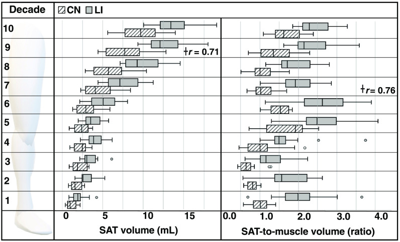
Patients with lipedema () and controls () matched for age and body mass index (BMI) underwent CSE-MRI acquired from the thighs to ankles. Images were segmented to partition SAT and skeletal muscle with a semiautomated algorithm incorporating classical image processing techniques (thresholding, active contours, Boolean operations, and morphological operations). The Dice similarity coefficient (DSC) was computed for SAT and muscle automated versus ground truth segmentations in the calf and thigh. SAT and muscle volumes and the SAT-to-muscle volume ratio were calculated across slices for decades containing 10% of total slices per participant. The effect size was calculated, and Mann-Whitney test applied to compare metrics in each decade between groups (significance: two-sided ).

RESULTS

Mean DSC for SAT segmentations was 0.96 in the calf and 0.98 in the thigh, and for muscle was 0.97 in the calf and 0.97 in the thigh. In all decades, mean SAT volume was significantly elevated in participants with versus without lipedema (), whereas muscle volume did not differ. Mean SAT-to-muscle volume ratio was significantly elevated () in all decades, where the greatest effect size for distinguishing lipedema was in the seventh decade approximately midthigh ().

CONCLUSIONS

The semiautomated segmentation of lower-extremity SAT and muscle from CSE-MRI could enable fast multislice analysis of SAT deposition throughout the legs relevant to distinguishing patients with lipedema from females with similar BMI but without SAT disease.

摘要

目的

脂肪性水肿是一种疼痛性皮下脂肪组织(SAT)疾病,其特征是下肢SAT不成比例地堆积,常被误诊为肥胖症。我们开发了一种半自动分割流程,用于从多层化学位移编码(CSE)磁共振成像(MRI)中量化脂肪性水肿患者独特的下肢SAT量。

方法

对年龄和体重指数(BMI)相匹配的脂肪性水肿患者()和对照组()进行从大腿到脚踝的CSE-MRI检查。采用结合经典图像处理技术(阈值处理、活动轮廓、布尔运算和形态学运算)的半自动算法对图像进行分割,以区分SAT和骨骼肌。计算小腿和大腿中SAT和肌肉自动分割与真实分割的骰子相似系数(DSC)。为每位参与者计算包含总切片10%的数十个切片的SAT和肌肉体积以及SAT与肌肉体积比。计算效应大小,并应用曼-惠特尼检验比较各组在每个十年中的指标(显著性:双侧)。

结果

SAT分割的平均DSC在小腿为0.96,在大腿为0.98;肌肉分割的平均DSC在小腿为0.97,在大腿为0.97。在所有十年中,有脂肪性水肿的参与者的平均SAT体积显著高于无脂肪性水肿的参与者(),而肌肉体积无差异。所有十年中的平均SAT与肌肉体积比均显著升高(),其中区分脂肪性水肿的最大效应大小在大约大腿中部的第七个十年()。

结论

从CSE-MRI中对下肢SAT和肌肉进行半自动分割,可以实现对整个腿部SAT沉积的快速多层分析,有助于区分脂肪性水肿患者与BMI相似但无SAT疾病的女性。

https://cdn.ncbi.nlm.nih.gov/pmc/blobs/bcd6/10185105/7bd8e8d5cec4/JMI-010-036001-g006.jpg
https://cdn.ncbi.nlm.nih.gov/pmc/blobs/bcd6/10185105/4bea22c57500/JMI-010-036001-g001.jpg
https://cdn.ncbi.nlm.nih.gov/pmc/blobs/bcd6/10185105/96c8f062ad26/JMI-010-036001-g002.jpg
https://cdn.ncbi.nlm.nih.gov/pmc/blobs/bcd6/10185105/7626763c59e5/JMI-010-036001-g003.jpg
https://cdn.ncbi.nlm.nih.gov/pmc/blobs/bcd6/10185105/841e492d0261/JMI-010-036001-g004.jpg
https://cdn.ncbi.nlm.nih.gov/pmc/blobs/bcd6/10185105/00f64f64ed13/JMI-010-036001-g005.jpg
https://cdn.ncbi.nlm.nih.gov/pmc/blobs/bcd6/10185105/7bd8e8d5cec4/JMI-010-036001-g006.jpg

相似文献

1
Semiautomated segmentation of lower extremity MRI reveals distinctive subcutaneous adipose tissue in lipedema: a pilot study.下肢MRI的半自动分割显示脂肪性水肿中独特的皮下脂肪组织:一项初步研究。
J Med Imaging (Bellingham). 2023 May;10(3):036001. doi: 10.1117/1.JMI.10.3.036001. Epub 2023 May 15.
2
Tissue Sodium Content is Elevated in the Skin and Subcutaneous Adipose Tissue in Women with Lipedema.脂肪水肿女性的皮肤和皮下脂肪组织中组织钠含量升高。
Obesity (Silver Spring). 2018 Feb;26(2):310-317. doi: 10.1002/oby.22090. Epub 2017 Dec 27.
3
Subcutaneous Adipose Tissue Edema in Lipedema Revealed by Noninvasive 3T MR Lymphangiography.非侵入性 3T MR 淋巴造影术显示脂肪性脂肪瘤中的皮下脂肪组织水肿。
J Magn Reson Imaging. 2023 Feb;57(2):598-608. doi: 10.1002/jmri.28281. Epub 2022 Jun 3.
4
Lipedema stage affects adipocyte hypertrophy, subcutaneous adipose tissue inflammation and interstitial fibrosis.脂肪水肿分期影响脂肪细胞肥大、皮下脂肪组织炎症和间质纤维化。
Front Immunol. 2023 Jul 28;14:1223264. doi: 10.3389/fimmu.2023.1223264. eCollection 2023.
5
Deep learning for standardized, MRI-based quantification of subcutaneous and subfascial tissue volume for patients with lipedema and lymphedema.深度学习在基于 MRI 的皮下和筋膜下组织容积标准化定量测量中的应用:用于脂肪水肿和淋巴水肿患者。
Eur Radiol. 2023 Feb;33(2):884-892. doi: 10.1007/s00330-022-09047-0. Epub 2022 Aug 17.
6
Pilot study: whole body manual subcutaneous adipose tissue (SAT) therapy improved pain and SAT structure in women with lipedema.试点研究:全身手动皮下脂肪组织(SAT)疗法改善了脂肪性水肿女性的疼痛和SAT结构。
Horm Mol Biol Clin Investig. 2017 Sep 20;33(2):/j/hmbci.2018.33.issue-2/hmbci-2017-0035/hmbci-2017-0035.xml. doi: 10.1515/hmbci-2017-0035.
7
Convolutional Neural Network-Based Automated Segmentation of Skeletal Muscle and Subcutaneous Adipose Tissue on Thigh MRI in Muscular Dystrophy Patients.基于卷积神经网络的肌营养不良患者大腿MRI上骨骼肌和皮下脂肪组织的自动分割
J Funct Morphol Kinesiol. 2024 Jul 12;9(3):123. doi: 10.3390/jfmk9030123.
8
Upper and Lower Extremity Measurement of Tissue Sodium and Fat Content in Patients with Lipedema.脂肪性水肿患者肢体组织钠和脂肪含量的测量。
Obesity (Silver Spring). 2020 May;28(5):907-915. doi: 10.1002/oby.22778. Epub 2020 Apr 9.
9
What is the best reference site for a single MRI slice to assess whole-body skeletal muscle and adipose tissue volumes in healthy adults?评估健康成年人全身骨骼肌和脂肪组织体积时,用于单个MRI切片的最佳参考网站是什么?
Am J Clin Nutr. 2015 Jul;102(1):58-65. doi: 10.3945/ajcn.115.111203. Epub 2015 May 27.
10
3D Chemical Shift-Encoded MRI for Volume and Composition Quantification of Abdominal Adipose Tissue During an Overfeeding Protocol in Healthy Volunteers.3D 化学位移编码 MRI 用于健康志愿者过食方案期间腹部脂肪组织容量和成分定量。
J Magn Reson Imaging. 2019 Jun;49(6):1587-1599. doi: 10.1002/jmri.26532. Epub 2018 Oct 16.

引用本文的文献

1
The effect of a low-carbohydrate diet on subcutaneous adipose tissue in females with lipedema.低碳水化合物饮食对患有脂肪性水肿女性皮下脂肪组织的影响。
Front Nutr. 2024 Nov 7;11:1484612. doi: 10.3389/fnut.2024.1484612. eCollection 2024.
2
Sex Differences in Fat Distribution and Muscle Fat Infiltration in the Lower Extremity: A Retrospective Diverse-Ethnicity 7T MRI Study in a Research Institute Setting in the USA.下肢脂肪分布和肌肉脂肪浸润的性别差异:美国一家研究机构的回顾性多民族7T MRI研究
Diagnostics (Basel). 2024 Oct 10;14(20):2260. doi: 10.3390/diagnostics14202260.

本文引用的文献

1
Subcutaneous Adipose Tissue Edema in Lipedema Revealed by Noninvasive 3T MR Lymphangiography.非侵入性 3T MR 淋巴造影术显示脂肪性脂肪瘤中的皮下脂肪组织水肿。
J Magn Reson Imaging. 2023 Feb;57(2):598-608. doi: 10.1002/jmri.28281. Epub 2022 Jun 3.
2
Label efficient segmentation of single slice thigh CT with two-stage pseudo labels.基于两阶段伪标签的单层面大腿CT图像的标签高效分割
J Med Imaging (Bellingham). 2022 Sep;9(5):052405. doi: 10.1117/1.JMI.9.5.052405. Epub 2022 May 19.
3
Persistent lipedema pain in patients after bariatric surgery: a case series of 13 patients.
肥胖症手术后患者持续存在脂性肥大疼痛:13 例病例系列。
Surg Obes Relat Dis. 2022 May;18(5):628-633. doi: 10.1016/j.soard.2021.12.027. Epub 2022 Jan 7.
4
Standard of care for lipedema in the United States.美国脂性水肿的护理标准。
Phlebology. 2021 Dec;36(10):779-796. doi: 10.1177/02683555211015887. Epub 2021 May 28.
5
Reproducibility and repeatability of MRI-based body composition analysis.基于磁共振成像的身体成分分析的可重复性和再现性
Magn Reson Med. 2020 Dec;84(6):3146-3156. doi: 10.1002/mrm.28360. Epub 2020 Jun 10.
6
Upper and Lower Extremity Measurement of Tissue Sodium and Fat Content in Patients with Lipedema.脂肪性水肿患者肢体组织钠和脂肪含量的测量。
Obesity (Silver Spring). 2020 May;28(5):907-915. doi: 10.1002/oby.22778. Epub 2020 Apr 9.
7
Lipedema: A Call to Action!脂肪水肿:行动起来!
Obesity (Silver Spring). 2019 Oct;27(10):1567-1576. doi: 10.1002/oby.22597.
8
MRI adipose tissue and muscle composition analysis-a review of automation techniques.MRI脂肪组织和肌肉成分分析——自动化技术综述
Br J Radiol. 2018 Sep;91(1089):20180252. doi: 10.1259/bjr.20180252. Epub 2018 Jul 24.
9
Tissue Sodium Content is Elevated in the Skin and Subcutaneous Adipose Tissue in Women with Lipedema.脂肪水肿女性的皮肤和皮下脂肪组织中组织钠含量升高。
Obesity (Silver Spring). 2018 Feb;26(2):310-317. doi: 10.1002/oby.22090. Epub 2017 Dec 27.
10
Lipedema: A Relatively Common Disease with Extremely Common Misconceptions.脂肪性水肿:一种相对常见但存在极为常见误解的疾病。
Plast Reconstr Surg Glob Open. 2016 Sep 28;4(9):e1043. doi: 10.1097/GOX.0000000000001043. eCollection 2016 Sep.