Department of Biomedicine, Aarhus University, Aarhus, Denmark.
Department of Surgery, Randers Regional Hospital, Randers, Denmark.
Int J Cancer. 2022 Oct 1;151(7):1150-1165. doi: 10.1002/ijc.34147. Epub 2022 Jun 17.
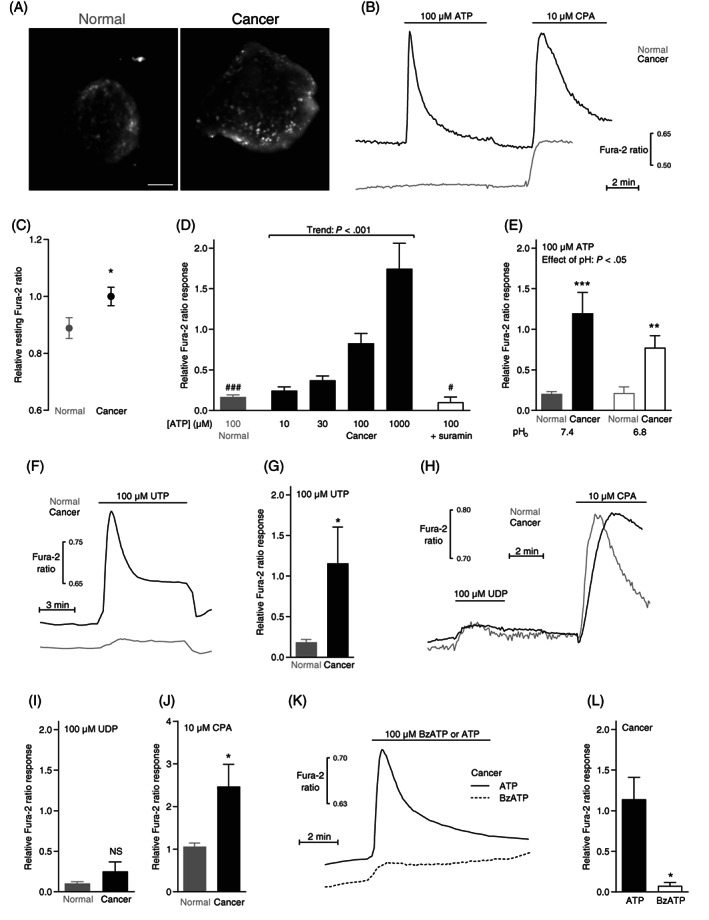
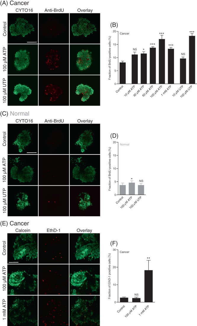
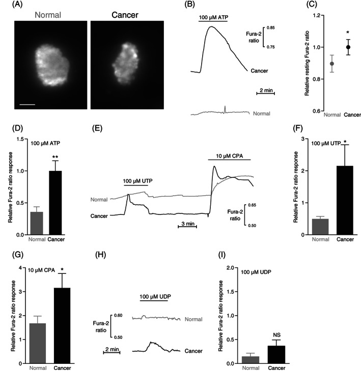
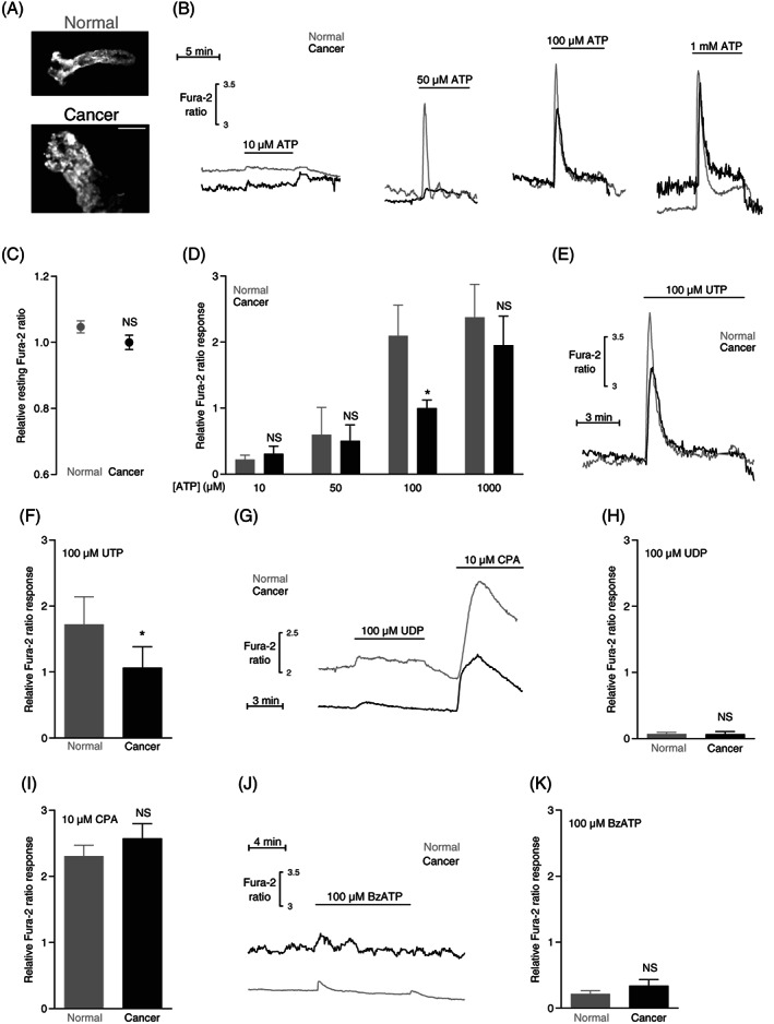
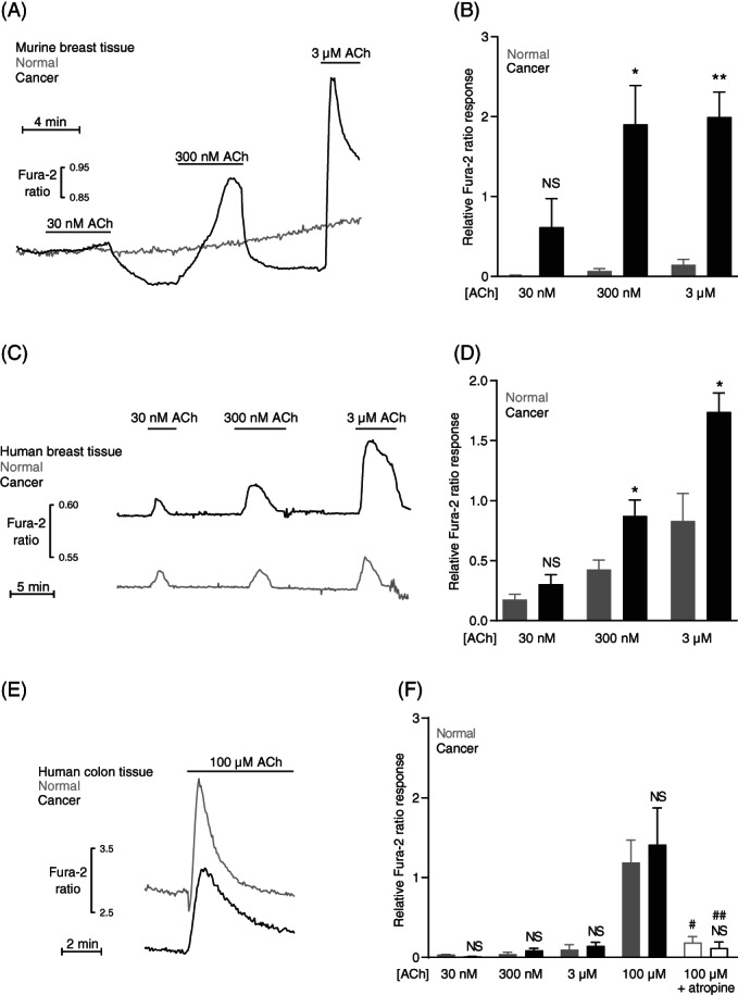
Intracellular Ca dynamics shape malignant behaviors of cancer cells. Whereas previous studies focused on cultured cancer cells, we here used breast organoids and colonic crypts freshly isolated from human and murine surgical biopsies. We performed fluorescence microscopy to evaluate intracellular Ca concentrations in breast and colon cancer tissue with preferential focus on intracellular Ca release in response to purinergic and cholinergic stimuli. Inhibition of the sarco-/endoplasmic reticulum Ca ATPase with cyclopiazonic acid elicited larger Ca responses in breast cancer tissue, but not in colon cancer tissue, relative to respective normal tissue. The resting intracellular Ca concentration was elevated, and ATP, UTP and acetylcholine induced strongly augmented intracellular Ca responses in breast cancer tissue compared with normal breast tissue. In contrast, resting intracellular Ca levels and acetylcholine-induced increases in intracellular Ca concentrations were unaffected and ATP- and UTP-induced Ca responses were smaller in colon cancer tissue compared with normal colon tissue. In accordance with the amplified Ca responses, ATP and UTP substantially increased proliferative activity-evaluated by bromodeoxyuridine incorporation-in breast cancer tissue, whereas the effect was minimal in normal breast tissue. ATP caused cell death-identified with ethidium homodimer-1 staining-in breast cancer tissue only at concentrations above the expected pathophysiological range. We conclude that intracellular Ca responses are amplified in breast cancer tissue, but not in colon cancer tissue, and that nucleotide signaling stimulates breast cancer cell proliferation within the extracellular concentration range typical for solid cancer tissue.
细胞内 Ca 动力学塑造了癌细胞的恶性行为。虽然之前的研究集中在培养的癌细胞上,但我们在这里使用了从人类和鼠手术活检中新鲜分离的乳腺类器官和结肠隐窝。我们通过荧光显微镜评估了乳腺癌和结肠癌组织中的细胞内 Ca 浓度,特别关注对嘌呤能和胆碱能刺激的细胞内 Ca 释放。与相应的正常组织相比,用环匹阿尼酸抑制肌浆/内质网 Ca-ATP 酶在乳腺癌组织中引起更大的 Ca 反应,但在结肠癌组织中没有。与正常乳腺组织相比,乳腺癌组织的静息细胞内 Ca 浓度升高,并且 ATP、UTP 和乙酰胆碱诱导的细胞内 Ca 反应强烈增强。相比之下,在结肠癌组织中,静息细胞内 Ca 水平和乙酰胆碱诱导的细胞内 Ca 浓度增加不受影响,而 ATP 和 UTP 诱导的 Ca 反应较小。与放大的 Ca 反应一致,ATP 和 UTP 显著增加了乳腺癌组织中的增殖活性(通过溴脱氧尿苷掺入评估),而在正常乳腺组织中则作用很小。只有在高于预期的病理生理范围的浓度下,ATP 才会导致乳腺癌组织中的细胞死亡(用 ethidium homodimer-1 染色鉴定)。我们得出结论,细胞内 Ca 反应在乳腺癌组织中被放大,但在结肠癌组织中没有被放大,核苷酸信号在典型的实体瘤组织细胞外浓度范围内刺激乳腺癌细胞增殖。