Voss Ninna C S, Dreyer Thomas, Henningsen Mikkel B, Vahl Pernille, Honoré Bent, Boedtkjer Ebbe
Department of Biomedicine, Aarhus University, DK-8000 Aarhus, Denmark.
Department of Surgery, Regionshospitalet Randers, DK-8930 Randers, Denmark.
Cancers (Basel). 2020 Apr 6;12(4):891. doi: 10.3390/cancers12040891.
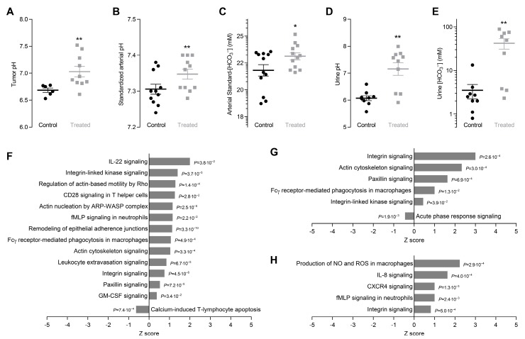
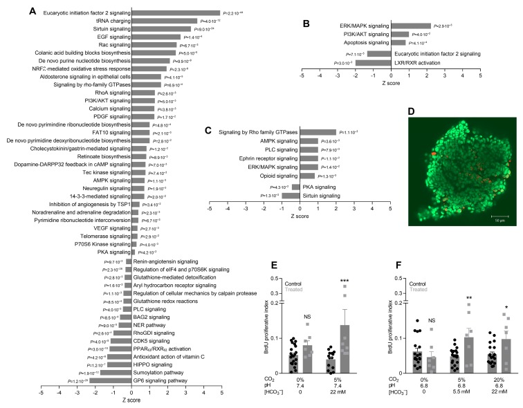
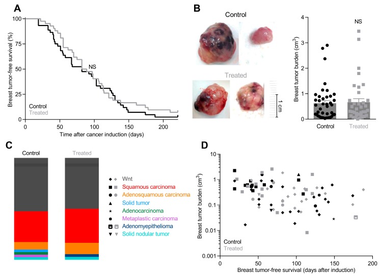
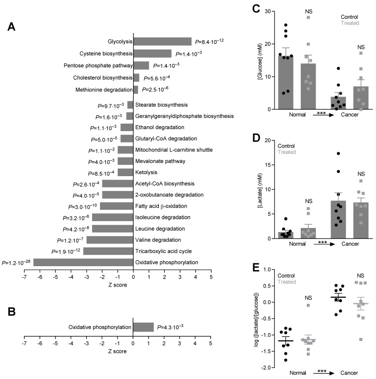
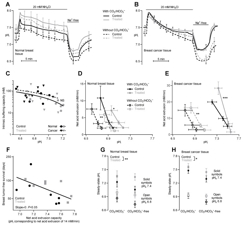
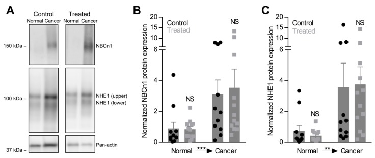
The acidic tumor microenvironment modifies malignant cell behavior. Here, we study consequences of the microenvironment in breast carcinomas. Beginning at carcinogen-based breast cancer induction, we supply either regular or NaHCO-containing drinking water to female C57BL/6j mice. We evaluate urine and blood acid-base status, tumor metabolism (microdialysis sampling), and tumor pH (pH-sensitive microelectrodes) in vivo. Based on freshly isolated epithelial organoids from breast carcinomas and normal breast tissue, we assess protein expression (immunoblotting, mass spectrometry), intracellular pH (fluorescence microscopy), and cell proliferation (bromodeoxyuridine incorporation). Oral NaHCO therapy increases breast tumor pH in vivo from 6.68 ± 0.04 to 7.04 ± 0.09 and intracellular pH in breast epithelial organoids by ~0.15. Breast tumors develop with median latency of 85.5 ± 8.2 days in NaHCO-treated mice vs. 82 ± 7.5 days in control mice. Oral NaHCO therapy does not affect tumor growth, histopathology or glycolytic metabolism. The capacity for cellular net acid extrusion is increased in NaHCO-treated mice and correlates negatively with breast tumor latency. Oral NaHCO therapy elevates proliferative activity in organoids from breast carcinomas. Changes in protein expression patterns-observed by high-throughput proteomics analyses-between cancer and normal breast tissue and in response to oral NaHCO therapy reveal complex influences on metabolism, cytoskeleton, cell-cell and cell-matrix interaction, and cell signaling pathways. We conclude that oral NaHCO therapy neutralizes the microenvironment of breast carcinomas, elevates the cellular net acid extrusion capacity, and accelerates proliferation without net effect on breast cancer development or tumor growth. We demonstrate unexpected pro-neoplastic consequences of oral NaHCO therapy that in breast tissue cancel out previously reported anti-neoplastic effects.
酸性肿瘤微环境会改变恶性细胞的行为。在此,我们研究乳腺癌微环境的影响。从基于致癌物诱导乳腺癌开始,我们为雌性C57BL/6j小鼠提供常规饮用水或含NaHCO₃的饮用水。我们在体内评估尿液和血液的酸碱状态、肿瘤代谢(微透析采样)以及肿瘤pH值(pH敏感微电极)。基于从乳腺癌和正常乳腺组织中新鲜分离的上皮类器官,我们评估蛋白质表达(免疫印迹、质谱分析)、细胞内pH值(荧光显微镜检查)以及细胞增殖(溴脱氧尿苷掺入)。口服NaHCO₃疗法可使体内乳腺癌肿瘤pH值从6.68±0.04升高至7.04±0.09,并使乳腺上皮类器官的细胞内pH值升高约0.15。在接受NaHCO₃治疗的小鼠中,乳腺癌发生的中位潜伏期为85.5±8.2天,而对照小鼠为82±7.5天。口服NaHCO₃疗法不影响肿瘤生长、组织病理学或糖酵解代谢。在接受NaHCO₃治疗的小鼠中,细胞净酸外排能力增强,且与乳腺癌潜伏期呈负相关。口服NaHCO₃疗法可提高乳腺癌类器官中的增殖活性。通过高通量蛋白质组学分析观察到的癌症与正常乳腺组织之间以及对口服NaHCO₃疗法反应中蛋白质表达模式的变化,揭示了对代谢、细胞骨架、细胞间和细胞与基质相互作用以及细胞信号通路的复杂影响。我们得出结论,口服NaHCO₃疗法可中和乳腺癌的微环境,提高细胞净酸外排能力,并加速增殖,但对乳腺癌发展或肿瘤生长无净影响。我们证明了口服NaHCO₃疗法意想不到的促肿瘤作用,即在乳腺组织中抵消了先前报道的抗肿瘤作用。