• 文献检索
  • 文档翻译
  • 深度研究
  • 学术资讯
  • Suppr Zotero 插件Zotero 插件
  • 邀请有礼
  • 套餐&价格
  • 历史记录
应用&插件
Suppr Zotero 插件Zotero 插件浏览器插件Mac 客户端Windows 客户端微信小程序
定价
高级版会员购买积分包购买API积分包
服务
文献检索文档翻译深度研究API 文档MCP 服务
关于我们
关于 Suppr公司介绍联系我们用户协议隐私条款
关注我们

Suppr 超能文献

核心技术专利:CN118964589B侵权必究
粤ICP备2023148730 号-1Suppr @ 2026

文献检索

告别复杂PubMed语法,用中文像聊天一样搜索,搜遍4000万医学文献。AI智能推荐,让科研检索更轻松。

立即免费搜索

文件翻译

保留排版,准确专业,支持PDF/Word/PPT等文件格式,支持 12+语言互译。

免费翻译文档

深度研究

AI帮你快速写综述,25分钟生成高质量综述,智能提取关键信息,辅助科研写作。

立即免费体验

蝶鞍和斜坡发育性、炎症性和肿瘤性病变的 CT 和 MRI 特征的实用概述。

A practical overview of CT and MRI features of developmental, inflammatory, and neoplastic lesions of the sphenoid body and clivus.

机构信息

Department of Experimental and Clinical Biomedical Sciences, Radiodiagnostic Unit N. 2, University of Florence - Azienda Ospedaliero-Universitaria Careggi, Largo Brambilla 3, 50134, Florence, Italy.

Department of Neuroradiology, Careggi University Hospital, Largo Piero Palagi 1, 50134, Florence, Italy.

出版信息

Neuroradiology. 2022 Aug;64(8):1483-1509. doi: 10.1007/s00234-022-02986-x. Epub 2022 Jun 3.

DOI:10.1007/s00234-022-02986-x
PMID:35657394
原文链接:https://pmc.ncbi.nlm.nih.gov/articles/PMC9271108/
Abstract

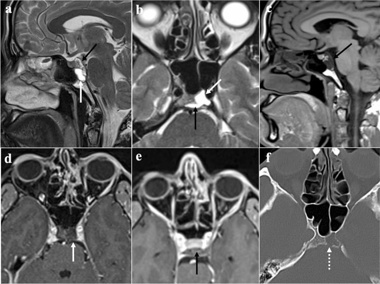
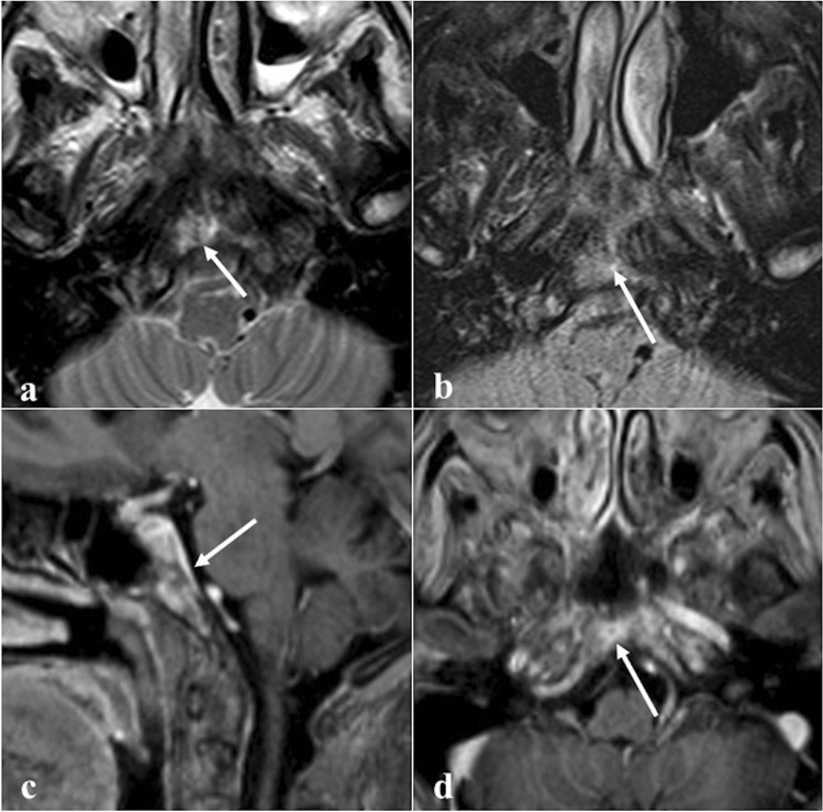
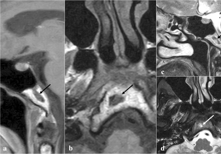
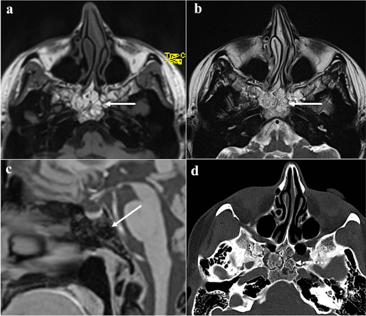
The sphenoid bone is an unpaired bone that contributes to the formation of the skull base. Despite the enormous progress in transnasal endoscopic visualisation, imaging techniques remain the cornerstones to characterise any pathological condition arising in this area. In the present review, we offer a bird's-eye view of the developmental, inflammatory, and neoplastic alterations affecting the sphenoid body and clivus, with the aim to propose a practical diagnostic aid for radiologists based on clinico-epidemiological, computed tomography, and magnetic resonance imaging features.

摘要

蝶骨是不成对的颅骨之一,参与颅底的形成。尽管经鼻内镜可视化技术取得了巨大进展,但影像学检查仍然是该区域任何病变特征的基石。在本综述中,我们对影响蝶骨体和斜坡的发育、炎症和肿瘤性改变进行了概述,旨在根据临床流行病学、计算机断层扫描和磁共振成像特征为放射科医生提供实用的诊断辅助。

https://cdn.ncbi.nlm.nih.gov/pmc/blobs/36f1/9271108/069eecfd9365/234_2022_2986_Fig19_HTML.jpg
https://cdn.ncbi.nlm.nih.gov/pmc/blobs/36f1/9271108/bf92306b917c/234_2022_2986_Fig1_HTML.jpg
https://cdn.ncbi.nlm.nih.gov/pmc/blobs/36f1/9271108/64d9404e16d7/234_2022_2986_Fig2_HTML.jpg
https://cdn.ncbi.nlm.nih.gov/pmc/blobs/36f1/9271108/868cfc69b994/234_2022_2986_Fig3_HTML.jpg
https://cdn.ncbi.nlm.nih.gov/pmc/blobs/36f1/9271108/88c20e9bad9c/234_2022_2986_Fig4_HTML.jpg
https://cdn.ncbi.nlm.nih.gov/pmc/blobs/36f1/9271108/0636c74c38fd/234_2022_2986_Fig5_HTML.jpg
https://cdn.ncbi.nlm.nih.gov/pmc/blobs/36f1/9271108/c29e5f84388f/234_2022_2986_Fig6_HTML.jpg
https://cdn.ncbi.nlm.nih.gov/pmc/blobs/36f1/9271108/7b11c43b73dc/234_2022_2986_Fig7_HTML.jpg
https://cdn.ncbi.nlm.nih.gov/pmc/blobs/36f1/9271108/9e02ceedef7a/234_2022_2986_Fig8_HTML.jpg
https://cdn.ncbi.nlm.nih.gov/pmc/blobs/36f1/9271108/e811ee182ca2/234_2022_2986_Fig9_HTML.jpg
https://cdn.ncbi.nlm.nih.gov/pmc/blobs/36f1/9271108/d5b5327aa95f/234_2022_2986_Fig10_HTML.jpg
https://cdn.ncbi.nlm.nih.gov/pmc/blobs/36f1/9271108/4204d541c7e5/234_2022_2986_Fig11_HTML.jpg
https://cdn.ncbi.nlm.nih.gov/pmc/blobs/36f1/9271108/5acd3cbbb7a0/234_2022_2986_Fig12_HTML.jpg
https://cdn.ncbi.nlm.nih.gov/pmc/blobs/36f1/9271108/c0fc53d35faa/234_2022_2986_Fig13_HTML.jpg
https://cdn.ncbi.nlm.nih.gov/pmc/blobs/36f1/9271108/69cfc502371d/234_2022_2986_Fig14_HTML.jpg
https://cdn.ncbi.nlm.nih.gov/pmc/blobs/36f1/9271108/95a9c3b347e7/234_2022_2986_Fig15_HTML.jpg
https://cdn.ncbi.nlm.nih.gov/pmc/blobs/36f1/9271108/3fbb6e425605/234_2022_2986_Fig16_HTML.jpg
https://cdn.ncbi.nlm.nih.gov/pmc/blobs/36f1/9271108/fc14858ce63c/234_2022_2986_Fig17_HTML.jpg
https://cdn.ncbi.nlm.nih.gov/pmc/blobs/36f1/9271108/6c90eb2945c5/234_2022_2986_Fig18_HTML.jpg
https://cdn.ncbi.nlm.nih.gov/pmc/blobs/36f1/9271108/069eecfd9365/234_2022_2986_Fig19_HTML.jpg

相似文献

1
A practical overview of CT and MRI features of developmental, inflammatory, and neoplastic lesions of the sphenoid body and clivus.蝶鞍和斜坡发育性、炎症性和肿瘤性病变的 CT 和 MRI 特征的实用概述。
Neuroradiology. 2022 Aug;64(8):1483-1509. doi: 10.1007/s00234-022-02986-x. Epub 2022 Jun 3.
2
Neuroimaging: intrinsic lesions of the central skull base region.神经影像学:中颅底区域的内在病变
Semin Ultrasound CT MR. 2013 Oct;34(5):412-35. doi: 10.1053/j.sult.2013.08.004.
3
Malignant giant cell tumor of the skull base originating from clivus and sphenoid bone.起源于斜坡和蝶骨的颅底恶性巨细胞瘤。
J Neurooncol. 2006 Jan;76(2):149-52. doi: 10.1007/s11060-005-4343-5.
4
Skull base erosion in nasopharyngeal carcinoma: detection by CT and MRI.鼻咽癌颅底骨质侵蚀:CT与MRI检测
Clin Radiol. 1996 Sep;51(9):625-31. doi: 10.1016/s0009-9260(96)80056-0.
5
A single-center experience with giant cell tumors of sphenoid bone and clivus.单发蝶骨和斜坡骨巨细胞瘤的单中心经验。
Tumori. 2021 Dec;107(6):NP94-NP100. doi: 10.1177/03008916211024357. Epub 2021 Aug 10.
6
Endoscopic approach to lesions of the sphenoid sinus, orbital apex, and clivus.经鼻内镜治疗蝶窦、眶尖和斜坡病变的方法。
Am J Otolaryngol. 2003 Sep-Oct;24(5):317-22. doi: 10.1016/s0196-0709(03)00062-0.
7
T classification and clivus margin as risk factors for determining locoregional control by radiotherapy of nasopharyngeal carcinoma.T分类和斜坡切缘作为鼻咽癌放疗确定局部区域控制的危险因素。
Cancer. 1998 Jan 15;82(2):261-7. doi: 10.1002/(sici)1097-0142(19980115)82:2<261::aid-cncr3>3.0.co;2-u.
8
Endoscopic transphenoidal approach for fibrous dysplasia of clivus, tuberculum sellae and sphenoid sinus; report of three cases.内镜经蝶入路治疗斜坡、鞍结节及蝶窦骨纤维异常增殖症:三例报告
Turk Neurosurg. 2012;22(5):662-6. doi: 10.5137/1019-5149.JTN.4060-10.0.
9
[Polyostotic Fibrous dysplasia of the clivus: atypical CT and MRI imaging].[斜坡多骨型骨纤维异常增殖症:非典型CT和MRI影像]
Rofo. 2013 Jan;185(1):77-9. doi: 10.1055/s-0032-1325341. Epub 2012 Oct 29.
10
[Endoscopic transnasal approach for surgical treatment of skull base clival area chordomas (Report of 2 cases)].[经鼻内镜入路手术治疗颅底斜坡区脊索瘤(附2例报告)]
Lin Chuang Er Bi Yan Hou Tou Jing Wai Ke Za Zhi. 2018 Feb;32(3):230-232. doi: 10.13201/j.issn.1001-1781.2018.03.019.

引用本文的文献

1
Rare Case of Metastatic Breast Cancer to the Sphenoid Sinus: Diagnosis and Treatment.蝶窦转移性乳腺癌罕见病例:诊断与治疗
Case Rep Oncol. 2025 Jul 8;18(1):1040-1049. doi: 10.1159/000546040. eCollection 2025 Jan-Dec.
2
Bilateral abducens nerve palsy caused by retroclival mass.斜坡后肿块导致双侧展神经麻痹。
Acta Neurol Belg. 2025 Mar 26. doi: 10.1007/s13760-025-02770-7.
3
Markedly T2-Hypointense Clival Plasmacytoma With Light Chain Deposition Disease: A Case Report.伴有轻链沉积病的明显T2低信号斜坡浆细胞瘤:一例报告

本文引用的文献

1
Arrested pneumatization of sinus sphenoid, revealed by hypo-acusis: A case report.听力减退提示蝶窦气化停滞:一例报告
Ann Med Surg (Lond). 2021 Oct 13;71:102939. doi: 10.1016/j.amsu.2021.102939. eCollection 2021 Nov.
2
The snowman sign in a patient with pituitary tumor apoplexy.垂体瘤卒中患者的雪人征。
J Community Hosp Intern Med Perspect. 2021 May 10;11(3):416-417. doi: 10.1080/20009666.2021.1898086. eCollection 2021.
3
The 2021 WHO Classification of Tumors of the Central Nervous System: a summary.2021 年世卫组织中枢神经系统肿瘤分类:概述。
Cureus. 2024 Dec 23;16(12):e76289. doi: 10.7759/cureus.76289. eCollection 2024 Dec.
4
A Rare Case of Benign Long-Standing Ecchordosis Physaliphora.一例罕见的长期存在的良性泡状脊索瘤病例。
Cureus. 2023 Nov 27;15(11):e49490. doi: 10.7759/cureus.49490. eCollection 2023 Nov.
5
Sphenoid sinus anatomical variants and pathologies: pictorial essay.蝶窦解剖变异与病变:影像学研究论文。
Neuroradiology. 2023 Aug;65(8):1187-1203. doi: 10.1007/s00234-023-03163-4. Epub 2023 May 18.
Neuro Oncol. 2021 Aug 2;23(8):1231-1251. doi: 10.1093/neuonc/noab106.
4
Magnetic resonance imaging of salivary gland tumours: Key findings for imaging characterisation.唾液腺肿瘤的磁共振成像:影像学特征的关键发现。
Eur J Radiol. 2021 Jun;139:109716. doi: 10.1016/j.ejrad.2021.109716. Epub 2021 Apr 10.
5
Chordoma-Current Understanding and Modern Treatment Paradigms.脊索瘤——当前的认识与现代治疗模式
J Clin Med. 2021 Mar 4;10(5):1054. doi: 10.3390/jcm10051054.
6
Diagnosis of Skull Base Osteomyelitis.颅底骨髓炎的诊断
Radiographics. 2021 Jan-Feb;41(1):156-174. doi: 10.1148/rg.2021200046.
7
Nelson Syndrome: Clival Invasion of Corticotroph Pituitary Adenoma Resulting in Alternating Sixth Nerve Palsies.纳尔逊综合征:促肾上腺皮质激素垂体腺瘤向鞍上侵袭导致动眼神经交替性麻痹。
J Neuroophthalmol. 2021 Mar 1;41(1):114-118. doi: 10.1097/WNO.0000000000001141.
8
Association Between Human Papilloma Virus Infection and Malignant Sinonasal Inverted Papilloma.人乳头瘤病毒感染与恶性鼻腔鼻窦内翻性乳头状瘤的关系。
Laryngoscope. 2021 Jun;131(6):1200-1205. doi: 10.1002/lary.29125. Epub 2020 Oct 28.
9
Sinonasal mucosal melanoma: a 10-year experience of 36 cases in China.鼻腔鼻窦黏膜黑色素瘤:中国36例患者的10年经验
Ann Transl Med. 2020 Aug;8(16):1022. doi: 10.21037/atm-20-5575.
10
Central skull base osteomyelitis secondary to invasive aspergillus sphenoid sinusitis presenting with isolated 12th nerve palsy.继发于侵袭性蝶窦曲霉菌病的中央颅底骨髓炎,表现为孤立性第12对脑神经麻痹。
IDCases. 2020 Aug 11;22:e00930. doi: 10.1016/j.idcr.2020.e00930. eCollection 2020.