Epithelial Systems Biology Laboratory, Systems Biology Center, National Heart, Lung, and Blood Institute, National Institutes of Health, Bethesda, MD, USA.
Cell Commun Signal. 2022 Jun 3;20(1):80. doi: 10.1186/s12964-022-00892-6.
A major goal in the discovery of cellular signaling networks is to identify regulated phosphorylation sites ("phosphosites") and map them to the responsible protein kinases. The V2 vasopressin receptor is a G-protein coupled receptor (GPCR) that is responsible for regulation of renal water excretion through control of aquaporin-2-mediated osmotic water transport in kidney collecting duct cells. Genome editing experiments have demonstrated that virtually all vasopressin-triggered phosphorylation changes are dependent on protein kinase A (PKA), but events downstream from PKA are still obscure.
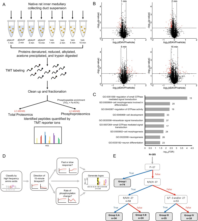
Here, we used: 1) Tandem mass tag-based quantitative phosphoproteomics to experimentally track phosphorylation changes over time in native collecting ducts isolated from rat kidneys; 2) a clustering algorithm to classify time course data based on abundance changes and the amino acid sequences surrounding the phosphosites; and 3) Bayes' Theorem to integrate the dynamic phosphorylation data with multiple prior "omic" data sets covering expression, subcellular location, known kinase activity, and characteristic surrounding sequences to identify a set of protein kinases that are regulated secondary to PKA activation.
Phosphoproteomic studies revealed 185 phosphosites regulated by vasopressin over 15 min. The resulting groups from the cluster algorithm were integrated with Bayes' Theorem to produce corresponding ranked lists of kinases likely responsible for each group. The top kinases establish three PKA-dependent protein kinase modules whose regulation mediate the physiological effects of vasopressin at a cellular level. The three modules are 1) a pathway involving several Rho/Rac/Cdc42-dependent protein kinases that control actin cytoskeleton dynamics; 2) mitogen-activated protein kinase and cyclin-dependent kinase pathways that control cell proliferation; and 3) calcium/calmodulin-dependent signaling.
Our findings identify a novel set of downstream small GTPase effectors and calcium/calmodulin-dependent kinases with potential roles in the regulation of water permeability through actin cytoskeleton rearrangement and aquaporin-2 trafficking. The proposed signaling network provides a stronger hypothesis for the kinases mediating V2 vasopressin receptor responses, encouraging future targeted examination via reductionist approaches. Furthermore, the Bayesian analysis described here provides a template for investigating signaling via other biological systems and GPCRs. Video abstract.
在细胞信号网络的发现中,一个主要目标是鉴定受调控的磷酸化位点(“磷酸化位点”),并将其映射到负责的蛋白激酶上。V2 血管加压素受体是一种 G 蛋白偶联受体(GPCR),通过控制肾脏集合管细胞中水通道蛋白 2 介导的渗透水转运,负责调节肾脏的水排泄。基因组编辑实验表明,几乎所有血管加压素触发的磷酸化变化都依赖于蛋白激酶 A(PKA),但 PKA 下游的事件仍不清楚。
在这里,我们使用了:1)基于串联质量标签的定量磷酸蛋白质组学,在从大鼠肾脏分离的天然集合管中,随时间推移实验性地跟踪磷酸化变化;2)聚类算法根据丰度变化和磷酸化位点周围的氨基酸序列对时间过程数据进行分类;3)贝叶斯定理将动态磷酸化数据与涵盖表达、亚细胞定位、已知激酶活性和特征周围序列的多个“组学”数据集相结合,以鉴定一组受 PKA 激活调节的蛋白激酶。
磷酸蛋白质组学研究揭示了血管加压素在 15 分钟内调节的 185 个磷酸化位点。聚类算法的结果与贝叶斯定理相结合,产生了相应的激酶排名列表,这些激酶可能对每个组负责。顶级激酶建立了三个 PKA 依赖性蛋白激酶模块,其调节介导了血管加压素在细胞水平上的生理效应。这三个模块是 1)涉及几种 Rho/Rac/Cdc42 依赖性蛋白激酶的途径,这些激酶控制肌动球蛋白细胞骨架动力学;2)有丝分裂原激活的蛋白激酶和细胞周期蛋白依赖性激酶途径,控制细胞增殖;3)钙/钙调蛋白依赖性信号转导。
我们的研究结果确定了一组新的下游小 GTPase 效应子和钙/钙调蛋白依赖性激酶,它们可能在通过肌动球蛋白细胞骨架重排和水通道蛋白 2 运输调节水通透性方面发挥作用。所提出的信号网络为介导 V2 血管加压素受体反应的激酶提供了一个更强的假说,鼓励未来通过还原论方法进行有针对性的检查。此外,本文描述的贝叶斯分析为研究其他生物系统和 GPCR 的信号转导提供了模板。视频摘要。