Department of Physiology and Pathophysiology, Rady Faculty of Health Sciences, University of Manitoba, Winnipeg, Canada; Children's Hospital Research Institute of Manitoba, 715 McDermot ave, Winnipeg, MB R3E 3P4, Canada.
Department of Physiology and Pathophysiology, Rady Faculty of Health Sciences, University of Manitoba, Winnipeg, Canada; Children's Hospital Research Institute of Manitoba, 715 McDermot ave, Winnipeg, MB R3E 3P4, Canada.
Mol Metab. 2022 Aug;62:101524. doi: 10.1016/j.molmet.2022.101524. Epub 2022 Jun 2.
Type 1 Diabetes (T1D) is characterized by progressive loss of insulin-producing pancreatic β cells as a result of autoimmune destruction. In addition to β cell death, recent work has shown that subpopulations of β cells acquire dysfunction during T1D. We previously reported that β cells undergoing a DNA damage response (DDR) and senescence accumulate during the pathogenesis of T1D. However, the question of how senescence develops in β cells has not been investigated.
Here, we tested the hypothesis that unrepaired DNA damage in the context of genetic susceptibility triggers β cell senescence using culture models including the mouse NIT1 β cell line derived from the T1D-susceptible nonobese diabetic (NOD) strain, human donor islets and EndoC β cells. DNA damage was chemically induced using etoposide or bleomycin and cells or islets were analyzed by a combination of molecular assays for senescence phenotypes including Western blotting, qRT-PCR, Luminex assays, flow cytometry and histochemical staining. RNA-seq was carried out to profile global transcriptomic changes in human islets undergoing DDR and senescence. Insulin ELISAs were used to quantify glucose-stimulated insulin secretion from chemically-induced senescent human islets, EndoC β cells and mouse β cell lines in culture.
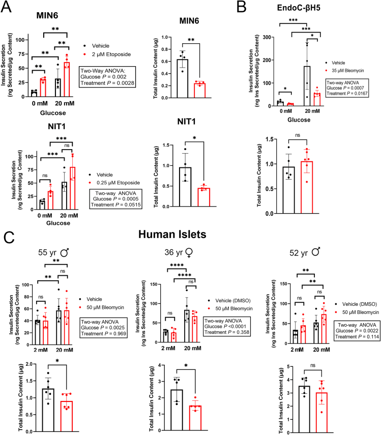
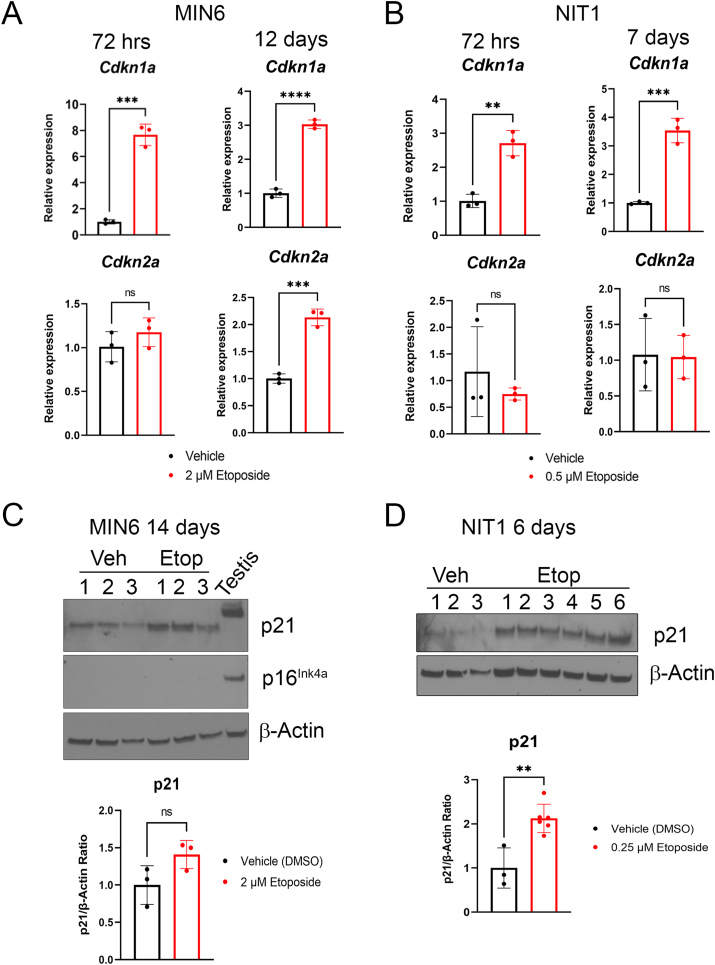
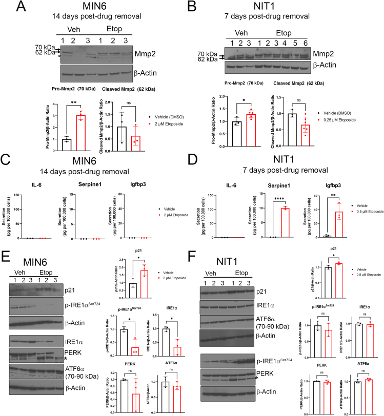
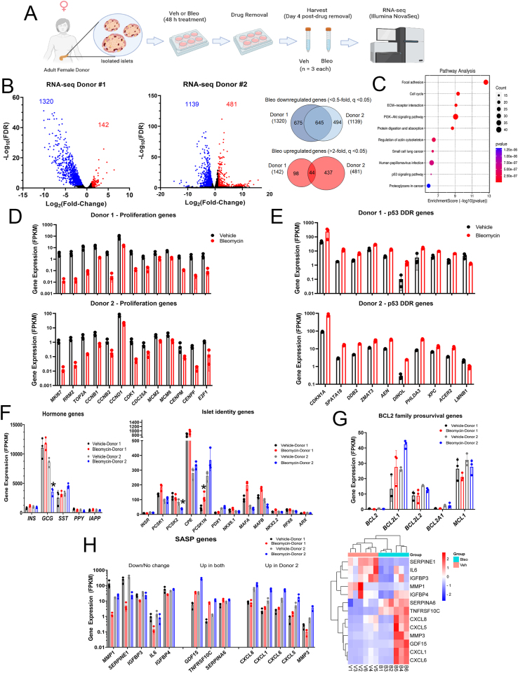
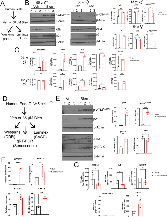
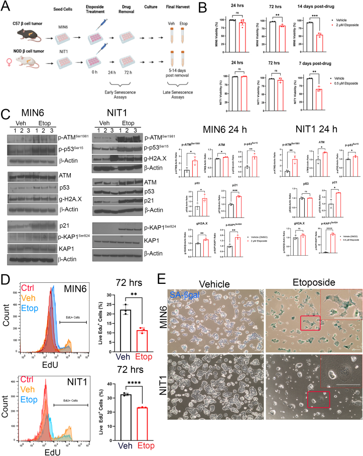
Sub-lethal DNA damage in NIT1 cells led to several classical hallmarks of senescence including sustained DDR activation, growth arrest, enlarged flattened morphology and a senescence-associated secretory phenotype (SASP) resembling what occurs in primary β cells during T1D in NOD mice. These phenotypes differed between NIT1 cells and the MIN6 β cell line derived from a non-T1D susceptible mouse strain. RNA-seq analysis of DNA damage-induced senescence in human islets from two different donors revealed a p53 transcriptional program and upregulation of prosurvival and SASP genes, with inter-donor variability in this response. Inter-donor variability in human islets was also apparent in the extent of persistent DDR activation and SASP at the protein level. Notably, chemically induced DNA damage also led to DDR activation and senescent phenotypes in EndoC-βH5 human β cells, confirming that this response can occur directly in a human β cell line. Finally, DNA damage led to different effects on glucose-stimulated insulin secretion in mouse β cell lines as compared with human islets and EndoC β cells.
Taken together, these findings suggest that some of the phenotypes of senescent β cells that accumulate during the development of T1D in the NOD mouse and humans can be modeled by chemically induced DNA damage to mouse β cell lines, human islets and EndoC β cells in culture. The differences between β cells from different mouse strains and different human islet donors and EndoC β cells highlights species differences and the role for genetic background in modifying the β cell response to DNA damage and its effects on insulin secretion. These culture models will be useful tools to understand some of the mechanisms of β cell senescence in T1D.
1 型糖尿病(T1D)的特征是由于自身免疫破坏导致产生胰岛素的胰腺β细胞进行性丧失。除了β细胞死亡外,最近的研究表明,在 T1D 发生过程中,β细胞亚群获得了功能障碍。我们之前报道过,在 T1D 发病过程中,经历 DNA 损伤反应(DDR)和衰老的β细胞会累积。然而,β细胞衰老如何发展的问题尚未得到研究。
在这里,我们通过使用包括来自 T1D 易感非肥胖型糖尿病(NOD)品系的小鼠 NIT1β细胞系、人供体胰岛和 EndoCβ细胞在内的培养模型,测试了在遗传易感性背景下未修复的 DNA 损伤引发β细胞衰老的假设。使用依托泊苷或博来霉素化学诱导 DNA 损伤,并用衰老表型的分子分析组合(包括 Western blot、qRT-PCR、Luminex 测定、流式细胞术和组织化学染色)来分析细胞或胰岛。进行 RNA-seq 以分析经历 DDR 和衰老的人胰岛中的全转录组变化。使用化学诱导的衰老人胰岛、EndoCβ细胞和小鼠β细胞系在培养中的葡萄糖刺激胰岛素分泌的 ELISA 进行定量。
NIT1 细胞中的亚致死性 DNA 损伤导致了几种衰老的典型特征,包括持续的 DDR 激活、生长停滞、扩大的扁平形态和类似于在 NOD 小鼠中发生 T1D 时原发性β细胞中发生的衰老相关分泌表型(SASP)。这些表型在 NIT1 细胞和来自非 T1D 易感小鼠品系的 MIN6β细胞系之间存在差异。来自两位不同供体的 DNA 损伤诱导的衰老人类胰岛的 RNA-seq 分析揭示了一个 p53 转录程序和促生存和 SASP 基因的上调,并且在这种反应中存在供体间的变异性。人类胰岛中的供体间变异性在 DDR 持续激活和蛋白质水平上的 SASP 方面也很明显。值得注意的是,化学诱导的 DNA 损伤也导致 EndoC-βH5 人类β细胞中 DDR 激活和衰老表型,证实这种反应可以直接在人类β细胞系中发生。最后,与人类胰岛和 EndoCβ细胞相比,DNA 损伤导致了不同的对葡萄糖刺激胰岛素分泌的影响小鼠β细胞系。
总之,这些发现表明,在 NOD 小鼠和人类中 T1D 发展过程中累积的一些衰老β细胞表型,可以通过化学诱导小鼠β细胞系、人类胰岛和 EndoCβ细胞中的 DNA 损伤来建模。来自不同小鼠品系和不同人类胰岛供体和 EndoCβ细胞的β细胞之间的差异突出了物种差异和遗传背景在修饰β细胞对 DNA 损伤的反应及其对胰岛素分泌的影响中的作用。这些培养模型将成为理解 T1D 中β细胞衰老的一些机制的有用工具。