• 文献检索
  • 文档翻译
  • 深度研究
  • 学术资讯
  • Suppr Zotero 插件Zotero 插件
  • 邀请有礼
  • 套餐&价格
  • 历史记录
应用&插件
Suppr Zotero 插件Zotero 插件浏览器插件Mac 客户端Windows 客户端微信小程序
定价
高级版会员购买积分包购买API积分包
服务
文献检索文档翻译深度研究API 文档MCP 服务
关于我们
关于 Suppr公司介绍联系我们用户协议隐私条款
关注我们

Suppr 超能文献

核心技术专利:CN118964589B侵权必究
粤ICP备2023148730 号-1Suppr @ 2026

文献检索

告别复杂PubMed语法,用中文像聊天一样搜索,搜遍4000万医学文献。AI智能推荐,让科研检索更轻松。

立即免费搜索

文件翻译

保留排版,准确专业,支持PDF/Word/PPT等文件格式,支持 12+语言互译。

免费翻译文档

深度研究

AI帮你快速写综述,25分钟生成高质量综述,智能提取关键信息,辅助科研写作。

立即免费体验

清除后的小鼠心脏中心肌细胞亚群的高分辨率三维心脏模型

High-Resolution 3D Heart Models of Cardiomyocyte Subpopulations in Cleared Murine Heart.

作者信息

Ren Huiying, Pu Zhaoli, Sun Tianyi, Chen Tangting, Liu Leiying, Liu Zhu, O'Shea Christopher, Pavlovic Davor, Tan Xiaoqiu, Lei Ming

机构信息

Laboratory of Medical Electrophysiology, Ministry of Education, Collaborative Innovation Center for Prevention and Treatment of Cardiovascular Disease/Institute of Cardiovascular Research, Luzhou Medical College, Luzhou, China.

Department of Cardiology, The Affiliated Hospital of Southwest Medical University, Luzhou, China.

出版信息

Front Physiol. 2022 May 18;13:779514. doi: 10.3389/fphys.2022.779514. eCollection 2022.

DOI:10.3389/fphys.2022.779514
PMID:35665220
原文链接:https://pmc.ncbi.nlm.nih.gov/articles/PMC9158482/
Abstract

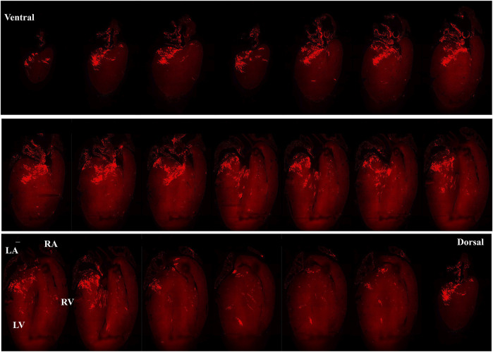
Biological tissues are naturally three-dimensional (3D) opaque structures, which poses a major challenge for the deep imaging of spatial distribution and localization of specific cell types in organs in biomedical research. Here we present a 3D heart imaging reconstruction approach by combining an improved heart tissue-clearing technique with high-resolution light-sheet fluorescence microscopy (LSFM). We have conducted a three-dimensional and multi-scale volumetric imaging of the ultra-thin planes of murine hearts for up to 2,000 images per heart in x-, y-, and z three directions. High-resolution 3D volume heart models were constructed in real-time by the Zeiss Zen program. By using such an approach, we investigated detailed three-dimensional spatial distributions of two specific cardiomyocyte populations including HCN4 expressing pacemaker cells and Pnmt cell-derived cardiomyocytes by using reporter mouse lines Hcn4 and Pnmt. HCN4 is distributed throughout right atrial nodal regions (i.e., sinoatrial and atrioventricular nodes) and the superior-inferior vena cava axis, while Pnmt cell-derived cardiomyocytes show distinct ventral, left heart, and dorsal side distribution pattern. Our further electrophysiological analysis indicates that Pnmt + cell-derived cardiomyocytes rich left ventricular (LV) base is more susceptible to ventricular arrhythmia under adrenergic stress than left ventricular apex or right ventricle regions. Thus, our 3D heart imaging reconstruction approach provides a new solution for studying the geometrical, topological, and physiological characteristics of specific cell types in organs.

摘要

生物组织是天然的三维(3D)不透明结构,这给生物医学研究中对器官内特定细胞类型的空间分布和定位进行深度成像带来了重大挑战。在此,我们提出一种3D心脏成像重建方法,该方法将改进的心脏组织透明化技术与高分辨率光片荧光显微镜(LSFM)相结合。我们对小鼠心脏的超薄平面进行了三维多尺度体积成像,每个心脏在x、y和z三个方向上最多可获取2000张图像。通过蔡司Zen程序实时构建了高分辨率3D心脏体积模型。利用这种方法,我们通过报告基因小鼠品系Hcn4和Pnmt研究了两种特定心肌细胞群体的详细三维空间分布,包括表达HCN4的起搏细胞和Pnmt细胞衍生的心肌细胞。HCN4分布于整个右心房结区(即窦房结和房室结)以及上下腔静脉轴,而Pnmt细胞衍生的心肌细胞呈现出明显的腹侧、左心和背侧分布模式。我们进一步的电生理分析表明,富含Pnmt + 细胞的左心室基部在肾上腺素能应激下比左心室尖部或右心室区域更容易发生室性心律失常。因此,我们的3D心脏成像重建方法为研究器官内特定细胞类型的几何、拓扑和生理特征提供了一种新的解决方案。

https://cdn.ncbi.nlm.nih.gov/pmc/blobs/322e/9158482/c142ca87d843/fphys-13-779514-g007.jpg
https://cdn.ncbi.nlm.nih.gov/pmc/blobs/322e/9158482/30a3673a5e8d/fphys-13-779514-g001.jpg
https://cdn.ncbi.nlm.nih.gov/pmc/blobs/322e/9158482/5403442723e2/fphys-13-779514-g002.jpg
https://cdn.ncbi.nlm.nih.gov/pmc/blobs/322e/9158482/6f0439836c25/fphys-13-779514-g003.jpg
https://cdn.ncbi.nlm.nih.gov/pmc/blobs/322e/9158482/40a096d57d7a/fphys-13-779514-g004.jpg
https://cdn.ncbi.nlm.nih.gov/pmc/blobs/322e/9158482/59dc38beb973/fphys-13-779514-g005.jpg
https://cdn.ncbi.nlm.nih.gov/pmc/blobs/322e/9158482/42c5e0373d5a/fphys-13-779514-g006.jpg
https://cdn.ncbi.nlm.nih.gov/pmc/blobs/322e/9158482/c142ca87d843/fphys-13-779514-g007.jpg

相似文献

1
High-Resolution 3D Heart Models of Cardiomyocyte Subpopulations in Cleared Murine Heart.清除后的小鼠心脏中心肌细胞亚群的高分辨率三维心脏模型
Front Physiol. 2022 May 18;13:779514. doi: 10.3389/fphys.2022.779514. eCollection 2022.
2
Optogenetic Control of Heart Rhythm by Selective Stimulation of Cardiomyocytes Derived from Pnmt Cells in Murine Heart.光遗传学控制源自小鼠心脏 Pnmt 细胞的心肌细胞的心率:选择性刺激。
Sci Rep. 2017 Jan 13;7:40687. doi: 10.1038/srep40687.
3
Three-dimensional image reconstruction of distribution of Pnmt cell-derived cells in murine heart.Pnmt 细胞源性细胞在小鼠心脏中分布的三维图像重建。
Sci Data. 2017 Sep 26;4:170134. doi: 10.1038/sdata.2017.134.
4
Three-dimensional structure of entire hydrated murine hearts at histological resolution.组织学分辨率下完整水合小鼠心脏的三维结构。
Sci Rep. 2025 Jan 22;15(1):2766. doi: 10.1038/s41598-024-83853-y.
5
Novel cardiac cell subpopulations: Pnmt-derived cardiomyocytes.新型心脏细胞亚群:Pnmt 衍生的心肌细胞。
Open Biol. 2020 Aug;10(8):200095. doi: 10.1098/rsob.200095. Epub 2020 Aug 19.
6
Innervation of sinoatrial nodal cardiomyocytes in mouse. A combined approach using immunofluorescent and electron microscopy.窦房结心肌细胞的神经支配。免疫荧光和电子显微镜联合应用的研究方法。
J Mol Cell Cardiol. 2014 Oct;75:188-97. doi: 10.1016/j.yjmcc.2014.07.016. Epub 2014 Aug 4.
7
Pnmt-Derived Cardiomyocytes: Anatomical Localization, Function and Future Perspectives.苯乙醇胺N-甲基转移酶衍生的心肌细胞:解剖定位、功能及未来展望
Front Physiol. 2019 Jul 10;10:713. doi: 10.3389/fphys.2019.00713. eCollection 2019.
8
Extended atrial conduction system characterised by the expression of the HCN4 channel and connexin45.以HCN4通道和连接蛋白45的表达为特征的延长心房传导系统。
Cardiovasc Res. 2006 Nov 1;72(2):271-81. doi: 10.1016/j.cardiores.2006.07.026. Epub 2006 Aug 2.
9
High-Speed Clearing and High-Resolution Staining for Analysis of Various Markers for Neurons and Vessels.高速清洗和高分辨率染色,用于分析神经元和血管的各种标志物。
Tissue Eng Regen Med. 2024 Oct;21(7):1037-1048. doi: 10.1007/s13770-024-00658-w. Epub 2024 Jul 2.
10
Distinctive left-sided distribution of adrenergic-derived cells in the adult mouse heart.成年小鼠心脏中去甲肾上腺素能衍生细胞的独特左侧分布。
PLoS One. 2011;6(7):e22811. doi: 10.1371/journal.pone.0022811. Epub 2011 Jul 27.

引用本文的文献

1
The role of cardiac pericytes in health and disease: therapeutic targets for myocardial infarction.心脏周细胞在健康与疾病中的作用:心肌梗死的治疗靶点
Nat Rev Cardiol. 2024 Feb;21(2):106-118. doi: 10.1038/s41569-023-00913-y. Epub 2023 Aug 4.

本文引用的文献

1
It's clearly the heart! Optical transparency, cardiac tissue imaging, and computer modelling.这显然是心脏!光学透明性、心脏组织成像和计算机建模。
Prog Biophys Mol Biol. 2022 Jan;168:18-32. doi: 10.1016/j.pbiomolbio.2021.06.005. Epub 2021 Jun 11.
2
Tissue clearing and imaging methods for cardiovascular development.用于心血管发育的组织透明化和成像方法。
iScience. 2021 Apr 1;24(4):102387. doi: 10.1016/j.isci.2021.102387. eCollection 2021 Apr 23.
3
Evidence of Superior and Inferior Sinoatrial Nodes in the Mammalian Heart.哺乳动物心脏中窦房结优势和劣势的证据。
JACC Clin Electrophysiol. 2020 Dec;6(14):1827-1840. doi: 10.1016/j.jacep.2020.09.012. Epub 2020 Nov 25.
4
Tissue clearing and its applications in neuroscience.组织透明化及其在神经科学中的应用。
Nat Rev Neurosci. 2020 Feb;21(2):61-79. doi: 10.1038/s41583-019-0250-1.
5
Advanced CUBIC tissue clearing for whole-organ cell profiling.高级 CUBIC 组织通透化技术用于整体器官细胞剖析。
Nat Protoc. 2019 Dec;14(12):3506-3537. doi: 10.1038/s41596-019-0240-9. Epub 2019 Nov 20.
6
Transcriptomic Profiling of the Developing Cardiac Conduction System at Single-Cell Resolution.转录组谱分析在单细胞分辨率下的心脏传导系统发育。
Circ Res. 2019 Aug 2;125(4):379-397. doi: 10.1161/CIRCRESAHA.118.314578. Epub 2019 Jul 9.
7
Whole-tissue biopsy phenotyping of three-dimensional tumours reveals patterns of cancer heterogeneity.三维肿瘤的全组织活检表型分析揭示了癌症异质性模式。
Nat Biomed Eng. 2017 Oct;1(10):796-806. doi: 10.1038/s41551-017-0139-0. Epub 2017 Oct 2.
8
Chemical Landscape for Tissue Clearing Based on Hydrophilic Reagents.基于亲水试剂的组织透明化化学全景图。
Cell Rep. 2018 Aug 21;24(8):2196-2210.e9. doi: 10.1016/j.celrep.2018.07.056.
9
An inhibitory antibody targeting carbonic anhydrase XII abrogates chemoresistance and significantly reduces lung metastases in an orthotopic breast cancer model in vivo.一种针对碳酸酐酶 XII 的抑制性抗体可消除化学耐药性,并在体内原位乳腺癌模型中显著减少肺转移。
Int J Cancer. 2018 Oct 15;143(8):2065-2075. doi: 10.1002/ijc.31607. Epub 2018 Aug 10.
10
3D Imaging and Quantitative Analysis of Vascular Networks: A Comparison of Ultramicroscopy and Micro-Computed Tomography.三维成像和血管网络定量分析:超微镜与微计算机断层扫描的比较。
Theranostics. 2018 Mar 7;8(8):2117-2133. doi: 10.7150/thno.22610. eCollection 2018.