Poflee Sandhya V, Bhatia Jasvinder Kaur
Department of Pathology, Goverment Medical College and Hospital, Nagpur, Maharashtra, India.
Department of Pathology, Command Hospital (Eastern Command), Kolkata, West Bengal, India.
Cytojournal. 2022 Apr 30;19:32. doi: 10.25259/CMAS_03_12_2021. eCollection 2022.
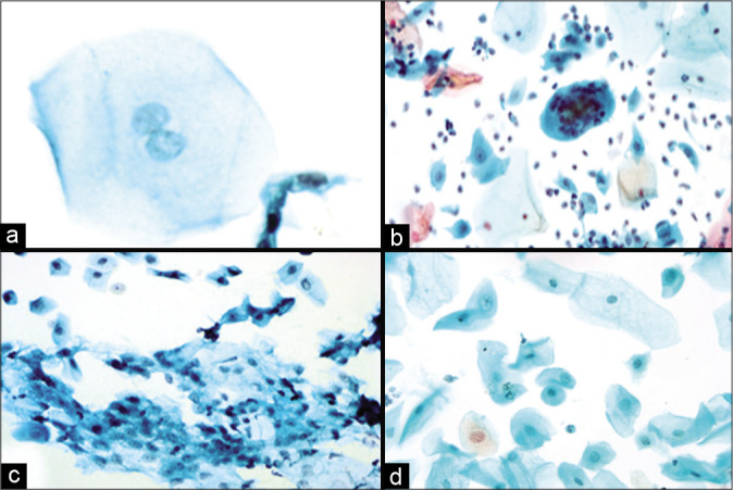
The different treatment options for carcinoma cervix include radiation, chemotherapy, and surgical treatments. Cytological analysis of smears is crucial for patient follow-up to determine response to therapy and to diagnose the persistence or recurrence of malignancy. Anatomical alterations and changes in cell morphology following radiation or chemotherapy make collecting and interpreting cervical cytology samples difficult. These issues can be mitigated by liquid-based cytology. Ionizing radiation is used in radiotherapy (RT) to kill cells. It is important that cytologists are aware of alterations in morphology of the cells. Radiation can cause cytoplasmic and nuclear changes. Cellular enlargement, vacuolation, granularity loss, and other changes linked with cell death are examples of cytoplasmic alterations. Nuclear enlargement and multinucleation are the most frequent nuclear alterations. These changes are determined by the amount of time that has passed since radiation. It should be emphasized that no one characteristic is pathognomonic. Post-irradiation dysplasia is a condition described as abnormal cellular changes in non-neoplastic epithelial cells after RT. Chemotherapy causes comparable alterations as radiation but impacts fewer cells. Busulfan and other chemotherapeutic treatments may produce morphological alterations, which cytologists must be aware of and able to identify. Immunosuppressive treatments, hormonal therapy, and tamoxifen are some of the other drugs that might cause changes in cervical morphology. Surgical methods used in the detection and treatment of cervical cancer may potentially cause alterations as a result of thermal damage and healing. For the treatment of cervical lesions, electrocautery and the loop electrosurgical excisional procedure are available. These procedures employ electric current ablation leading to ischemic changes in the cervical smear. Cytological analysis of smears following treatment with these modalities necessitates a comprehensive history, kind of therapy, and duration of treatment.
宫颈癌的不同治疗选择包括放疗、化疗和手术治疗。涂片的细胞学分析对于患者随访至关重要,以确定对治疗的反应并诊断恶性肿瘤的持续或复发。放疗或化疗后解剖结构的改变和细胞形态的变化使得收集和解读宫颈细胞学样本变得困难。液基细胞学可以缓解这些问题。电离辐射用于放射治疗(RT)以杀死细胞。细胞学家了解细胞形态的改变很重要。辐射可导致细胞质和细胞核的变化。细胞肿大、空泡化、颗粒丧失以及与细胞死亡相关的其他变化是细胞质改变的例子。核肿大和多核化是最常见的细胞核改变。这些变化取决于放疗后的时间长短。应该强调的是,没有一种特征是具有诊断意义的。放疗后发育异常是指放疗后非肿瘤性上皮细胞出现异常细胞变化的一种情况。化疗引起的改变与放疗类似,但影响的细胞较少。白消安和其他化疗治疗可能会产生形态学改变,细胞学家必须了解并能够识别这些改变。免疫抑制治疗、激素治疗和他莫昔芬是其他一些可能导致宫颈形态改变的药物。用于检测和治疗宫颈癌的手术方法可能由于热损伤和愈合而潜在地导致改变。对于宫颈病变的治疗,可以采用电灼术和环形电切术。这些手术采用电流消融,导致宫颈涂片出现缺血性改变。对接受这些治疗方式后的涂片进行细胞学分析需要全面了解病史、治疗类型和治疗持续时间。