• 文献检索
  • 文档翻译
  • 深度研究
  • 学术资讯
  • Suppr Zotero 插件Zotero 插件
  • 邀请有礼
  • 套餐&价格
  • 历史记录
应用&插件
Suppr Zotero 插件Zotero 插件浏览器插件Mac 客户端Windows 客户端微信小程序
定价
高级版会员购买积分包购买API积分包
服务
文献检索文档翻译深度研究API 文档MCP 服务
关于我们
关于 Suppr公司介绍联系我们用户协议隐私条款
关注我们

Suppr 超能文献

核心技术专利:CN118964589B侵权必究
粤ICP备2023148730 号-1Suppr @ 2026

文献检索

告别复杂PubMed语法,用中文像聊天一样搜索,搜遍4000万医学文献。AI智能推荐,让科研检索更轻松。

立即免费搜索

文件翻译

保留排版,准确专业,支持PDF/Word/PPT等文件格式,支持 12+语言互译。

免费翻译文档

深度研究

AI帮你快速写综述,25分钟生成高质量综述,智能提取关键信息,辅助科研写作。

立即免费体验

肿瘤相关巨噬细胞激活的 CD44 信号通路诱导腔面乳腺癌中细胞的集体脱落。

Activation of CD44 signaling in leader cells induced by tumor-associated macrophages drives collective detachment in luminal breast carcinomas.

机构信息

Department of Clinical Laboratory, Shanghai Jiao Tong University Affiliated Sixth People's Hospital, Shanghai, 200233, China.

Department of Molecular Biology Laboratory, Shanghai Jiao Tong University Affiliated Sixth People's Hospital, Shanghai, 200233, China.

出版信息

Cell Death Dis. 2022 Jun 9;13(6):540. doi: 10.1038/s41419-022-04986-4.

DOI:10.1038/s41419-022-04986-4
PMID:35680853
原文链接:https://pmc.ncbi.nlm.nih.gov/articles/PMC9184589/
Abstract

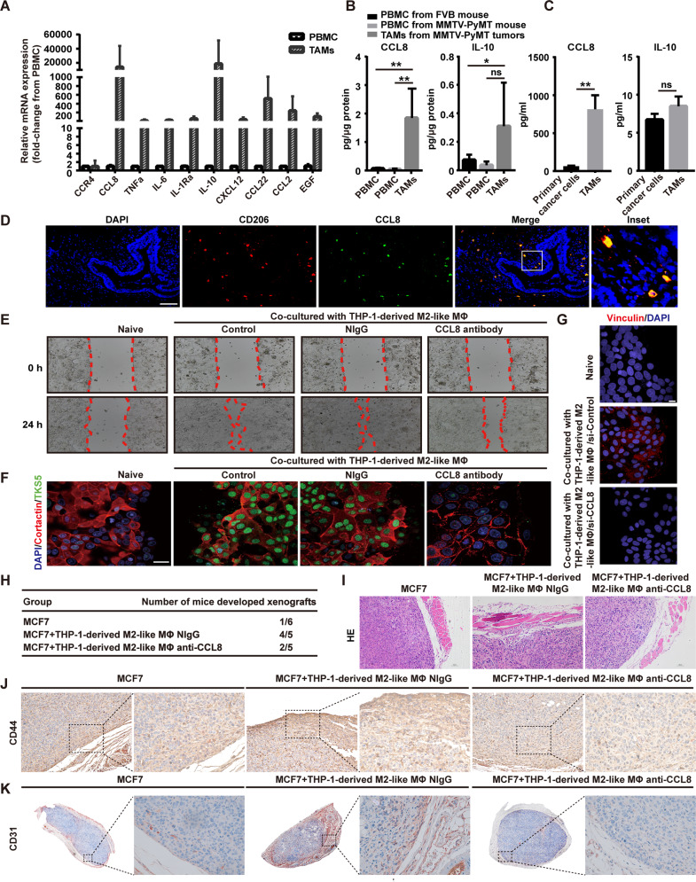
Collective detachment of cancer cells at the invading front could generate efficient metastatic spread. However, how cancer cell clusters shed from the leading front remains unknown. We previously reported that the dynamic expression of CD44 in breast cancers (BrCas) at collectively invading edges was associated with tumor-associated macrophages (TAMs). In this study, we first observed that the highly expressed CD44 (CD44) cancer cell clusters were located in the BrCa circulating vessels, accompanied by CD206 TAMs. Next, we identified that the cancer cell clusters can be converted to an invasive CD44 state which was induced by TAMs, thus giving rise to CD44-associated signaling mediated cohesive detachment. Then, we showed that disrupting CD44-signaling inhibited the TAMs triggered cohesive detaching using 3D organotypic culture and mouse models. Furthermore, our mechanistic study showed that the acquisition of CD44 state was mediated by the MDM2/p53 pathway activation which was induced by CCL8 released from TAMs. Blocking of CCL8 could inhibit the signaling cascade which decreased the CD44-mediated cohesive detachment and spread. Our findings uncover a novel mechanism underlying collective metastasis in BrCas that may be helpful to seek for potential targets.

摘要

癌细胞在侵袭前沿的集体脱离可能会产生有效的转移扩散。然而,癌细胞簇如何从前沿脱落仍然未知。我们之前的研究报告表明,在集体侵袭边缘的乳腺癌(BrCas)中 CD44 的动态表达与肿瘤相关巨噬细胞(TAMs)有关。在这项研究中,我们首先观察到,高表达 CD44(CD44)的癌细胞簇位于 BrCa 循环血管中,伴有 CD206 TAMs。接下来,我们确定了癌细胞簇可以转化为一种侵袭性的 CD44 状态,这种状态是由 TAMs 诱导的,从而导致 CD44 相关信号介导的粘着性脱离。然后,我们通过 3D 器官培养和小鼠模型表明,破坏 CD44 信号可以抑制 TAMs 触发的粘着性脱离。此外,我们的机制研究表明,CCL8 从 TAMs 释放后激活 MDM2/p53 通路,从而介导 CD44 状态的获得。阻断 CCL8 可以抑制信号级联反应,从而减少 CD44 介导的粘着性脱离和扩散。我们的研究结果揭示了 BrCas 中集体转移的一个新机制,这可能有助于寻找潜在的靶点。

https://cdn.ncbi.nlm.nih.gov/pmc/blobs/8921/9184589/3861bc45592b/41419_2022_4986_Fig6_HTML.jpg
https://cdn.ncbi.nlm.nih.gov/pmc/blobs/8921/9184589/8f194ab3df75/41419_2022_4986_Fig1_HTML.jpg
https://cdn.ncbi.nlm.nih.gov/pmc/blobs/8921/9184589/982d35bdbab3/41419_2022_4986_Fig2_HTML.jpg
https://cdn.ncbi.nlm.nih.gov/pmc/blobs/8921/9184589/972144711667/41419_2022_4986_Fig3_HTML.jpg
https://cdn.ncbi.nlm.nih.gov/pmc/blobs/8921/9184589/5bb7cf2e8ec3/41419_2022_4986_Fig4_HTML.jpg
https://cdn.ncbi.nlm.nih.gov/pmc/blobs/8921/9184589/cc833012f1af/41419_2022_4986_Fig5_HTML.jpg
https://cdn.ncbi.nlm.nih.gov/pmc/blobs/8921/9184589/3861bc45592b/41419_2022_4986_Fig6_HTML.jpg

相似文献

1
Activation of CD44 signaling in leader cells induced by tumor-associated macrophages drives collective detachment in luminal breast carcinomas.肿瘤相关巨噬细胞激活的 CD44 信号通路诱导腔面乳腺癌中细胞的集体脱落。
Cell Death Dis. 2022 Jun 9;13(6):540. doi: 10.1038/s41419-022-04986-4.
2
Inducible formation of leader cells driven by CD44 switching gives rise to collective invasion and metastases in luminal breast carcinomas.诱导性的 CD44 开关驱动的前体细胞形成导致腔面乳腺癌的集体侵袭和转移。
Oncogene. 2019 Nov;38(46):7113-7132. doi: 10.1038/s41388-019-0899-y. Epub 2019 Aug 15.
3
Cancer Cell CD44 Mediates Macrophage/Monocyte-Driven Regulation of Head and Neck Cancer Stem Cells.肿瘤细胞 CD44 介导巨噬细胞/单核细胞对头颈部癌症干细胞的调控作用。
Cancer Res. 2020 Oct 1;80(19):4185-4198. doi: 10.1158/0008-5472.CAN-20-1079. Epub 2020 Aug 14.
4
CD44/HA signaling mediates acquired resistance to a PI3Kα inhibitor.CD44/HA 信号转导介导对 PI3Kα 抑制剂获得性耐药。
Cell Death Dis. 2020 Oct 6;11(10):831. doi: 10.1038/s41419-020-03037-0.
5
CD44 enhances invasion of basal-like breast cancer cells by upregulating serine protease and collagen-degrading enzymatic expression and activity.CD44 通过上调丝氨酸蛋白酶和胶原降解酶的表达和活性增强基底样乳腺癌细胞的侵袭。
Breast Cancer Res. 2012 May 23;14(3):R84. doi: 10.1186/bcr3199.
6
Homophilic CD44 Interactions Mediate Tumor Cell Aggregation and Polyclonal Metastasis in Patient-Derived Breast Cancer Models.同质型 CD44 相互作用介导患者来源的乳腺癌模型中的肿瘤细胞聚集和多克隆转移。
Cancer Discov. 2019 Jan;9(1):96-113. doi: 10.1158/2159-8290.CD-18-0065. Epub 2018 Oct 25.
7
CD44 attenuates metastatic invasion during breast cancer progression.CD44在乳腺癌进展过程中减弱转移侵袭。
Cancer Res. 2005 Aug 1;65(15):6755-63. doi: 10.1158/0008-5472.CAN-05-0863.
8
The hyaluronan receptors CD44 and Rhamm (CD168) form complexes with ERK1,2 that sustain high basal motility in breast cancer cells.透明质酸受体CD44和Rhamm(CD168)与ERK1、2形成复合物,维持乳腺癌细胞的高基础运动性。
J Biol Chem. 2007 Jun 1;282(22):16667-80. doi: 10.1074/jbc.M702078200. Epub 2007 Mar 28.
9
CD44 activation state regulated by the CD44v10 isoform determines breast cancer proliferation.CD44v10 异构体调控的 CD44 激活状态决定乳腺癌的增殖。
Oncol Rep. 2021 Apr;45(4). doi: 10.3892/or.2021.7958. Epub 2021 Mar 2.
10
Breast Cancer Cell-Derived Soluble CD44 Promotes Tumor Progression by Triggering Macrophage IL1β Production.乳腺癌细胞衍生的可溶性 CD44 通过触发巨噬细胞 IL1β 产生促进肿瘤进展。
Cancer Res. 2020 Mar 15;80(6):1342-1356. doi: 10.1158/0008-5472.CAN-19-2288. Epub 2020 Jan 22.

引用本文的文献

1
Biomarkers, isolation methods, and therapeutic implications of breast cancer stem cells.乳腺癌干细胞的生物标志物、分离方法及治疗意义
Cancer Pathog Ther. 2025 Jan 23;3(5):392-401. doi: 10.1016/j.cpt.2025.01.006. eCollection 2025 Sep.
2
TGF-β signaling redirects Sox11 gene regulatory activity to promote partial EMT and collective invasion of oncogenically transformed intestinal organoids.转化生长因子-β(TGF-β)信号传导重定向Sox11基因调控活性,以促进致癌转化的肠道类器官的部分上皮-间质转化和集体侵袭。
Oncogenesis. 2025 May 20;14(1):17. doi: 10.1038/s41389-025-00560-7.
3
scSDNE: A semi-supervised method for inferring cell-cell interactions based on graph embedding.

本文引用的文献

1
Endometrial epithelial cells-derived exosomes deliver microRNA-30c to block the BCL9/Wnt/CD44 signaling and inhibit cell invasion and migration in ovarian endometriosis.子宫内膜上皮细胞衍生的外泌体传递微小RNA-30c以阻断BCL9/ Wnt/ CD44信号通路,并抑制卵巢子宫内膜异位症中的细胞侵袭和迁移。
Cell Death Discov. 2022 Apr 2;8(1):151. doi: 10.1038/s41420-022-00941-6.
2
The membrane receptor CD44: novel insights into metabolism.膜受体CD44:对新陈代谢的新见解
Trends Endocrinol Metab. 2022 May;33(5):318-332. doi: 10.1016/j.tem.2022.02.002. Epub 2022 Mar 4.
3
Prognostic Significance and Gene Co-Expression Network of and in Gliomas.
scSDNE:一种基于图嵌入推断细胞间相互作用的半监督方法。
PLoS Comput Biol. 2025 May 7;21(5):e1013027. doi: 10.1371/journal.pcbi.1013027. eCollection 2025 May.
4
Integrated multi-omics analysis reveals the functional and prognostic significance of lactylation-related gene PRDX1 in breast cancer.综合多组学分析揭示了乳酰化相关基因PRDX1在乳腺癌中的功能及预后意义。
Front Mol Biosci. 2025 Apr 4;12:1580622. doi: 10.3389/fmolb.2025.1580622. eCollection 2025.
5
Tumor cell-derived hyaluronan fragments induce endocytosis of S1PR1 to promote lymphangiogenesis through LYVE-1-Src pathway.肿瘤细胞衍生的透明质酸片段通过LYVE-1-Src途径诱导S1PR1内吞以促进淋巴管生成。
J Cancer. 2025 Jan 27;16(5):1466-1478. doi: 10.7150/jca.104309. eCollection 2025.
6
MDM2 inhibitors in cancer immunotherapy: Current status and perspective.MDM2抑制剂在癌症免疫治疗中的现状与展望
Genes Dis. 2024 Mar 28;11(6):101279. doi: 10.1016/j.gendis.2024.101279. eCollection 2024 Nov.
7
The inhibition of pancreatic cancer progression by K-Ras-overexpressing mesenchymal stem cell-derived secretomes.K-Ras 过表达间充质干细胞来源的分泌组抑制胰腺癌进展。
Sci Rep. 2023 Sep 12;13(1):15036. doi: 10.1038/s41598-023-41835-6.
胶质瘤中[具体基因名称]的预后意义及基因共表达网络
Front Oncol. 2022 Jan 11;11:602321. doi: 10.3389/fonc.2021.602321. eCollection 2021.
4
Long non-coding RNA AFAP1-AS1 accelerates lung cancer cells migration and invasion by interacting with SNIP1 to upregulate c-Myc.长链非编码 RNA AFAP1-AS1 通过与 SNIP1 相互作用来上调 c-Myc,从而促进肺癌细胞的迁移和侵袭。
Signal Transduct Target Ther. 2021 Jun 25;6(1):240. doi: 10.1038/s41392-021-00562-y.
5
PLAU Promotes Cell Proliferation and Epithelial-Mesenchymal Transition in Head and Neck Squamous Cell Carcinoma.PLAU促进头颈部鳞状细胞癌的细胞增殖和上皮-间质转化。
Front Genet. 2021 May 20;12:651882. doi: 10.3389/fgene.2021.651882. eCollection 2021.
6
Macrophage-tumor cell intertwine drives the transition into a mesenchymal-like cellular state of glioblastoma.肿瘤细胞与巨噬细胞相互交织促使胶质母细胞瘤向间充质样细胞状态转变。
Cancer Cell. 2021 Jun 14;39(6):743-745. doi: 10.1016/j.ccell.2021.05.003. Epub 2021 Jun 3.
7
Redirecting macrophage function to sustain their "defender" antitumor activity.将巨噬细胞功能重定向以维持其“防御者”抗肿瘤活性。
Cancer Cell. 2021 Jun 14;39(6):734-737. doi: 10.1016/j.ccell.2021.03.002. Epub 2021 Apr 1.
8
Regulation of Collective Metastasis by Nanolumenal Signaling.纳米腔室信号调控集体转移。
Cell. 2020 Oct 15;183(2):395-410.e19. doi: 10.1016/j.cell.2020.08.045. Epub 2020 Oct 1.
9
Monocyte-derived macrophages promote breast cancer bone metastasis outgrowth.单核细胞来源的巨噬细胞促进乳腺癌骨转移的生长。
J Exp Med. 2020 Nov 2;217(11). doi: 10.1084/jem.20191820.
10
Remodelling of the bone marrow microenvironment by stromal hyaluronan modulates the malignancy of breast cancer cells.基质透明质酸重塑骨髓微环境调节乳腺癌细胞的恶性程度。
Cell Commun Signal. 2020 Jun 9;18(1):89. doi: 10.1186/s12964-020-00592-z.