Noguchi Takaaki, Hirao Makoto, Tsuji Shigeyoshi, Etani Yuki, Ebina Kosuke, Tsuboi Hideki, Okamura Gensuke, Akita Shosuke, Okada Seiji, Hashimoto Jun
Orthopaedic Surgery, National Hospital Organization, Osaka Minami Medical Center, Kawachinagano, JPN.
Orthopaedic Surgery, Nippon Life Hospital, Osaka, JPN.
Cureus. 2022 May 8;14(5):e24831. doi: 10.7759/cureus.24831. eCollection 2022 May.
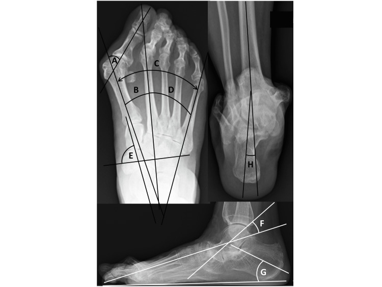
Increasing of intermetatarsal angle between the first and second metatarsals (M1-M2A) has been reported as a risk factor for recurrence of hallux valgus (HV) deformity, on the other hand, increasing of intermetatarsal angle between the second and fifth metatarsals (M2-M5A) has been reported as a risk factor for resubluxation of the metatarsophalangeal (MTP) joint of the lesser toe after rheumatoid forefoot surgery. In this study, parameters related to increasing M2-M5A were investigated, as compared with M1-M2A and M1-M5A.
Radiographic parameters including M1-M2A, M1-M5A, and M2-M5A were retrospectively evaluated for 119 lower limbs from 68 patients with rheumatoid arthritis (RA). To clarify the clinical importance of these intermetatarsal angles, relationships with results from the timed up-and-go (TUG) test were also investigated.
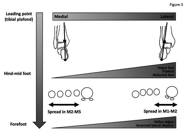
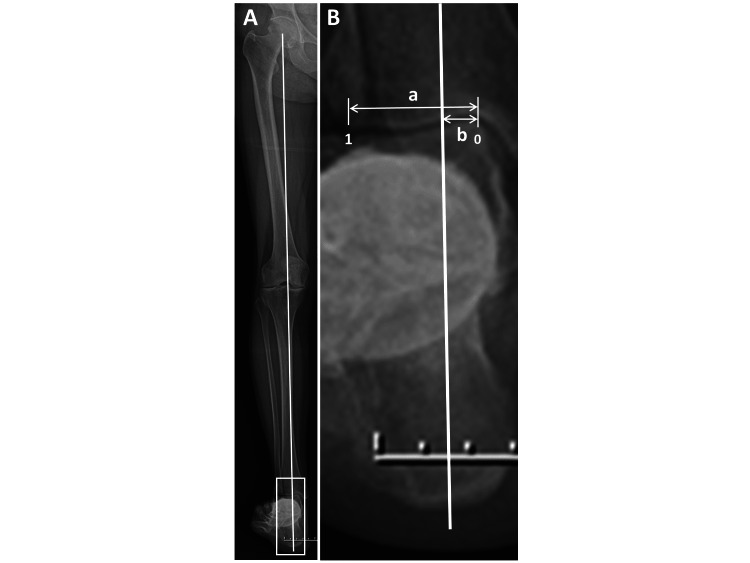
M1-M5A showed no correlation with mid-hind foot parameters, whereas M1-M2A and M2-M5A correlated with valgus/varus parameters. An increased M1-M2A was associated with lateral shift of the loading axis in the tibial plafond, whereas an increased M2-M5A was associated with medial shift, but M1-M5A showed no associations. M2-M5A/M1-M2A was significantly lower (1.7) in the normal TUG group than in the delayed TUG group (2.8) (p=0.045).
Different patterns of spread are seen for the forefoot. One has a predominantly increased M1-M2A with lateral shift of the loading point in the tibial plafond, whereas the other has a predominantly increased M2-M5A with medial shift of the loading point in the tibial plafond. M2-M5A also should be calculated, and M2-M5A/M1-M2A might be meaningful in understanding physical mobility in RA patients.
据报道,第一和第二跖骨间角(M1-M2A)增大是拇外翻(HV)畸形复发的危险因素,另一方面,第二和第五跖骨间角(M2-M5A)增大被报道为类风湿性前足手术后小趾跖趾(MTP)关节半脱位的危险因素。在本研究中,与M1-M2A和M1-M5A相比,对与M2-M5A增大相关的参数进行了研究。
对68例类风湿关节炎(RA)患者的119条下肢进行回顾性评估,包括M1-M2A、M1-M5A和M2-M5A等影像学参数。为了阐明这些跖骨间角的临床重要性,还研究了它们与定时起立行走(TUG)试验结果的关系。
M1-M5A与中后足参数无相关性,而M1-M2A和M2-M5A与外翻/内翻参数相关。M1-M2A增大与胫骨平台负重轴的外侧移位有关,而M2-M5A增大与内侧移位有关,但M1-M5A无相关性。正常TUG组的M2-M5A/M1-M2A显著低于延迟TUG组(分别为1.7和2.8)(p = 0.045)。
前足呈现不同的扩展模式。一种主要是M1-M2A增大,同时胫骨平台负重点向外侧移位;另一种主要是M2-M5A增大,同时胫骨平台负重点向内侧移位。还应计算M2-M5A,M2-M5A/M1-M2A可能有助于理解RA患者的身体活动能力。