• 文献检索
  • 文档翻译
  • 深度研究
  • 学术资讯
  • Suppr Zotero 插件Zotero 插件
  • 邀请有礼
  • 套餐&价格
  • 历史记录
应用&插件
Suppr Zotero 插件Zotero 插件浏览器插件Mac 客户端Windows 客户端微信小程序
定价
高级版会员购买积分包购买API积分包
服务
文献检索文档翻译深度研究API 文档MCP 服务
关于我们
关于 Suppr公司介绍联系我们用户协议隐私条款
关注我们

Suppr 超能文献

核心技术专利:CN118964589B侵权必究
粤ICP备2023148730 号-1Suppr @ 2026

文献检索

告别复杂PubMed语法,用中文像聊天一样搜索,搜遍4000万医学文献。AI智能推荐,让科研检索更轻松。

立即免费搜索

文件翻译

保留排版,准确专业,支持PDF/Word/PPT等文件格式,支持 12+语言互译。

免费翻译文档

深度研究

AI帮你快速写综述,25分钟生成高质量综述,智能提取关键信息,辅助科研写作。

立即免费体验

Eclipse Smart Segmentation 与 MIM Atlas Segment 行钇-90 选择性内放射治疗肝脏勾画的比较。

Comparison of Eclipse Smart Segmentation and MIM Atlas Segment for liver delineation for yttrium-90 selective internal radiation therapy.

机构信息

Department of Radiation Oncology, Thomas Jefferson University, Philadelphia, Pennsylvania, USA.

出版信息

J Appl Clin Med Phys. 2022 Aug;23(8):e13668. doi: 10.1002/acm2.13668. Epub 2022 Jun 15.

DOI:10.1002/acm2.13668
PMID:35702944
原文链接:https://pmc.ncbi.nlm.nih.gov/articles/PMC9359022/
Abstract

PURPOSE

The aim was to compare Smart Segmentation of Eclipse treatment planning system and Atlas Segment of MIM software for liver delineation for resin yttrium-90 (Y-90) procedures.

MATERIALS AND METHODS

CT images of 20 patients treated with resin Y-90 selective internal radiation therapy (SIRT) were tested. Liver contours generated with Smart Segmentation and Atlas Segment were compared with physician manually delineated contours. Dice similarity coefficient (DSC), mean distance to agreement (MDA), and ratio of volume (RV) were calculated. The contours were evaluated with activity calculations and ratio of activity (RA) was calculated.

RESULTS

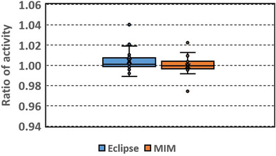
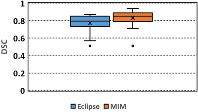
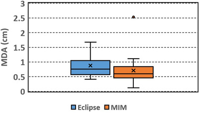
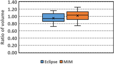
Mean DSCs were 0.77 and 0.83, MDAs were 0.88 and 0.71 cm, mean RVs were 0.95 and 1.02, and mean RAs were 1.00 and 1.00, for Eclipse and MIM results, respectively.

CONCLUSION

MIM outperformed Eclipse in both DSC and MDA, whereas the differences in liver volumes and calculated activities were statistically insignificant between the Eclipse and MIM results. Both auto-segmentation tools can be used to generate initial liver contours for resin Y-90 SIRT, which need to be reviewed and edited by physicians.

摘要

目的

比较 Eclipse 治疗计划系统的 Smart Segmentation 和 MIM 软件的 Atlas Segment 进行钇-90(Y-90)树脂治疗的肝脏勾画。

材料和方法

对 20 例接受树脂 Y-90 选择性内放射治疗(SIRT)的患者的 CT 图像进行了测试。将 Smart Segmentation 和 Atlas Segment 生成的肝轮廓与医生手动勾画的轮廓进行比较。计算了 Dice 相似系数(DSC)、平均一致性距离(MDA)和体积比(RV)。对轮廓进行了活性计算,并计算了活性比(RA)。

结果

Eclipse 和 MIM 的平均 DSC 分别为 0.77 和 0.83,MDA 分别为 0.88 和 0.71cm,平均 RV 分别为 0.95 和 1.02,平均 RA 分别为 1.00 和 1.00。

结论

在 DSC 和 MDA 方面,MIM 优于 Eclipse,而 Eclipse 和 MIM 结果之间肝体积和计算活性的差异无统计学意义。这两种自动分割工具都可以用于生成钇-90 树脂 SIRT 的初始肝轮廓,这些轮廓需要由医生进行审查和编辑。

https://cdn.ncbi.nlm.nih.gov/pmc/blobs/f54a/9359022/ce247f5cd7d8/ACM2-23-e13668-g005.jpg
https://cdn.ncbi.nlm.nih.gov/pmc/blobs/f54a/9359022/347888517a40/ACM2-23-e13668-g002.jpg
https://cdn.ncbi.nlm.nih.gov/pmc/blobs/f54a/9359022/5ffd7df2abf9/ACM2-23-e13668-g004.jpg
https://cdn.ncbi.nlm.nih.gov/pmc/blobs/f54a/9359022/170b8e7e37a8/ACM2-23-e13668-g003.jpg
https://cdn.ncbi.nlm.nih.gov/pmc/blobs/f54a/9359022/6503e8d181dd/ACM2-23-e13668-g001.jpg
https://cdn.ncbi.nlm.nih.gov/pmc/blobs/f54a/9359022/ce247f5cd7d8/ACM2-23-e13668-g005.jpg

相似文献

1
Comparison of Eclipse Smart Segmentation and MIM Atlas Segment for liver delineation for yttrium-90 selective internal radiation therapy.Eclipse Smart Segmentation 与 MIM Atlas Segment 行钇-90 选择性内放射治疗肝脏勾画的比较。
J Appl Clin Med Phys. 2022 Aug;23(8):e13668. doi: 10.1002/acm2.13668. Epub 2022 Jun 15.
2
Evaluation of Atlas-based auto-segmentation of liver in MR images for Yttrium-90 selective internal radiation therapy.基于图谱的磁共振图像肝脏自动分割在钇-90 选择性内放射治疗中的评价。
J Appl Clin Med Phys. 2023 May;24(5):e13979. doi: 10.1002/acm2.13979. Epub 2023 Apr 17.
3
Comparison of commercial atlas-based automatic segmentation software for prostate radiotherapy treatment planning.商业图谱自动分割软件在前列腺放射治疗计划中的比较。
Phys Eng Sci Med. 2024 Sep;47(3):881-894. doi: 10.1007/s13246-024-01411-2. Epub 2024 Apr 22.
4
Augmenting atlas-based liver segmentation for radiotherapy treatment planning by incorporating image features proximal to the atlas contours.通过合并靠近图谱轮廓的图像特征来增强基于图谱的肝脏分割用于放射治疗计划。
Phys Med Biol. 2017 Jan 7;62(1):272-288. doi: 10.1088/1361-6560/62/1/272. Epub 2016 Dec 17.
5
Auto-segmentation of low-risk clinical target volume for head and neck radiation therapy.头颈部放射治疗中低危临床靶区的自动勾画。
Pract Radiat Oncol. 2014 Jan-Feb;4(1):e31-7. doi: 10.1016/j.prro.2013.03.003. Epub 2013 May 3.
6
Comparative clinical evaluation of atlas and deep-learning-based auto-segmentation of organ structures in liver cancer.基于 atlas 和深度学习的肝癌器官结构自动分割的临床对比评估。
Radiat Oncol. 2019 Nov 27;14(1):213. doi: 10.1186/s13014-019-1392-z.
7
Clinical validation of an automatic atlas-based segmentation tool for male pelvis CT images.基于图谱的自动分割工具对男性骨盆 CT 图像的临床验证。
J Appl Clin Med Phys. 2022 Mar;23(3):e13507. doi: 10.1002/acm2.13507. Epub 2022 Jan 22.
8
Clinical feasibility of deep learning-based auto-segmentation of target volumes and organs-at-risk in breast cancer patients after breast-conserving surgery.保乳手术后乳腺癌患者基于深度学习的靶区体积和危及器官自动分割的临床可行性
Radiat Oncol. 2021 Feb 25;16(1):44. doi: 10.1186/s13014-021-01771-z.
9
Evaluation of a commercial DIR platform for contour propagation in prostate cancer patients treated with IMRT/VMAT.评价一个商业的 DIR 平台在接受调强放疗/VMAT 治疗的前列腺癌患者中的靶区勾画。
J Appl Clin Med Phys. 2020 Feb;21(2):14-25. doi: 10.1002/acm2.12787.
10
Methodological approach to create an atlas using a commercial auto-contouring software.使用商业自动勾画软件创建图谱的方法学方法。
J Appl Clin Med Phys. 2020 Dec;21(12):219-230. doi: 10.1002/acm2.13093. Epub 2020 Nov 25.

引用本文的文献

1
Dosimetric study on radioembolization with 166Ho poly L-lactic acid microspheres: dead time effects on prediction power.166Ho聚L-乳酸微球放射性栓塞的剂量学研究:死时间对预测能力的影响。
EJNMMI Phys. 2025 Jul 3;12(1):64. doi: 10.1186/s40658-025-00779-8.
2
Deep Learning-Based Auto-Segmentation for Liver Yttrium-90 Selective Internal Radiation Therapy.基于深度学习的肝脏钇-90选择性内放射治疗自动分割
Technol Cancer Res Treat. 2025 Jan-Dec;24:15330338251327081. doi: 10.1177/15330338251327081. Epub 2025 Mar 28.
3
Oncologic Applications of Artificial Intelligence and Deep Learning Methods in CT Spine Imaging-A Systematic Review.

本文引用的文献

1
Methodological approach to create an atlas using a commercial auto-contouring software.使用商业自动勾画软件创建图谱的方法学方法。
J Appl Clin Med Phys. 2020 Dec;21(12):219-230. doi: 10.1002/acm2.13093. Epub 2020 Nov 25.
2
Automated detection and delineation of hepatocellular carcinoma on multiphasic contrast-enhanced MRI using deep learning.使用深度学习在多期对比增强磁共振成像上自动检测和勾画肝细胞癌
Abdom Radiol (NY). 2021 Jan;46(1):216-225. doi: 10.1007/s00261-020-02604-5. Epub 2020 Jun 4.
3
Atlas-based auto-segmentation for postoperative radiotherapy planning in endometrial and cervical cancers.
人工智能和深度学习方法在CT脊柱成像中的肿瘤学应用——一项系统综述
Cancers (Basel). 2024 Aug 28;16(17):2988. doi: 10.3390/cancers16172988.
4
Comparison of commercial atlas-based automatic segmentation software for prostate radiotherapy treatment planning.商业图谱自动分割软件在前列腺放射治疗计划中的比较。
Phys Eng Sci Med. 2024 Sep;47(3):881-894. doi: 10.1007/s13246-024-01411-2. Epub 2024 Apr 22.
5
Prospective deployment of an automated implementation solution for artificial intelligence translation to clinical radiation oncology.人工智能翻译自动化实施解决方案在临床放射肿瘤学中的前瞻性部署。
Front Oncol. 2024 Jan 4;13:1305511. doi: 10.3389/fonc.2023.1305511. eCollection 2023.
6
Evaluation of manual and automated approaches for segmentation and extraction of quantitative indices from [F]FDG PET-CT images.评估手动和自动方法从 [F]FDG PET-CT 图像中分割和提取定量指标。
Biomed Phys Eng Express. 2024 Jan 5;10(2):025007. doi: 10.1088/2057-1976/ad160e.
7
A deep learning segmentation method to assess dose to organs at risk during breast radiotherapy.一种用于评估乳腺癌放疗期间危及器官剂量的深度学习分割方法。
Phys Imaging Radiat Oncol. 2023 Nov 21;28:100520. doi: 10.1016/j.phro.2023.100520. eCollection 2023 Oct.
8
Evaluation of Atlas-based auto-segmentation of liver in MR images for Yttrium-90 selective internal radiation therapy.基于图谱的磁共振图像肝脏自动分割在钇-90 选择性内放射治疗中的评价。
J Appl Clin Med Phys. 2023 May;24(5):e13979. doi: 10.1002/acm2.13979. Epub 2023 Apr 17.
基于图谱的自动分割在子宫内膜癌和宫颈癌术后放疗计划中的应用。
Radiat Oncol. 2020 May 13;15(1):106. doi: 10.1186/s13014-020-01562-y.
4
Efficacy evaluation of 2D, 3D U-Net semantic segmentation and atlas-based segmentation of normal lungs excluding the trachea and main bronchi.二维、三维 U-Net 语义分割与基于图谱的气管和主支气管外正常肺分割的效能评估。
J Radiat Res. 2020 Mar 23;61(2):257-264. doi: 10.1093/jrr/rrz086.
5
Clinical Evaluation of Commercial Atlas-Based Auto-Segmentation in the Head and Neck Region.头颈部区域基于图谱的商用自动分割的临床评估
Front Oncol. 2019 Apr 9;9:239. doi: 10.3389/fonc.2019.00239. eCollection 2019.
6
Fully automatic liver segmentation combining multi-dimensional graph cut with shape information in 3D CT images.三维 CT 图像中结合多维图割与形状信息的全自动肝脏分割。
Sci Rep. 2018 Jul 16;8(1):10700. doi: 10.1038/s41598-018-28787-y.
7
Automatic Intracranial Segmentation: Is the Clinician Still Needed?自动颅内分割:临床医生还有必要吗?
Technol Cancer Res Treat. 2018 Jan 1;17:1533034617748839. doi: 10.1177/1533034617748839.
8
Comparison of Automated Atlas-Based Segmentation Software for Postoperative Prostate Cancer Radiotherapy.基于图谱的自动分割软件在前列腺癌术后放疗中的比较
Front Oncol. 2016 Aug 3;6:178. doi: 10.3389/fonc.2016.00178. eCollection 2016.
9
Atlas-based liver segmentation and hepatic fat-fraction assessment for clinical trials.基于图谱的肝脏分割和肝脂肪分数评估用于临床试验。
Comput Med Imaging Graph. 2015 Apr;41:80-92. doi: 10.1016/j.compmedimag.2014.05.012. Epub 2014 Jun 9.
10
Liver Selective Internal Radiation Therapy with (90)Y resin microspheres: comparison between pre-treatment activity calculation methods.使用(90)Y树脂微球的肝脏选择性内放射治疗:治疗前活度计算方法的比较
Phys Med. 2014 Nov;30(7):752-64. doi: 10.1016/j.ejmp.2014.05.004. Epub 2014 Jun 9.