Department of Medicine, School of Clinical Sciences at Monash Health, Centre for Inflammatory Diseases, Monash Medical Centre, Monash University, Clayton, VIC, Australia.
Immunol Cell Biol. 2022 Aug;100(7):482-496. doi: 10.1111/imcb.12560. Epub 2022 Jun 15.
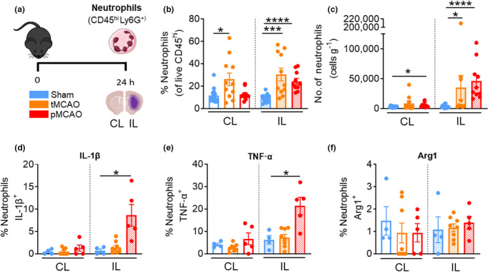
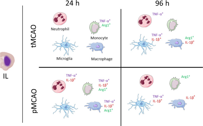
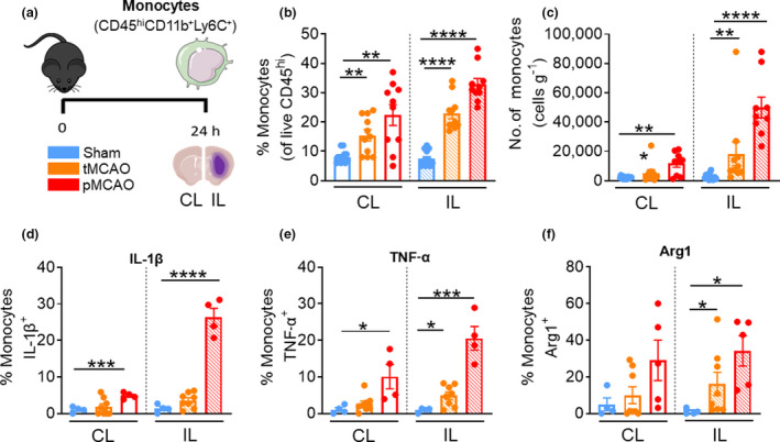
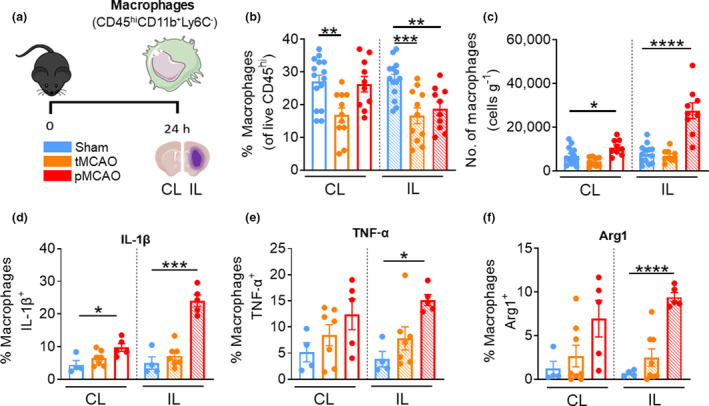
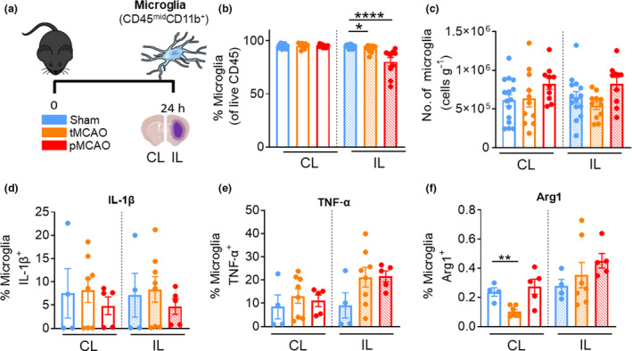
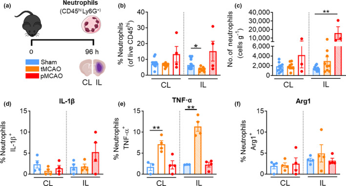
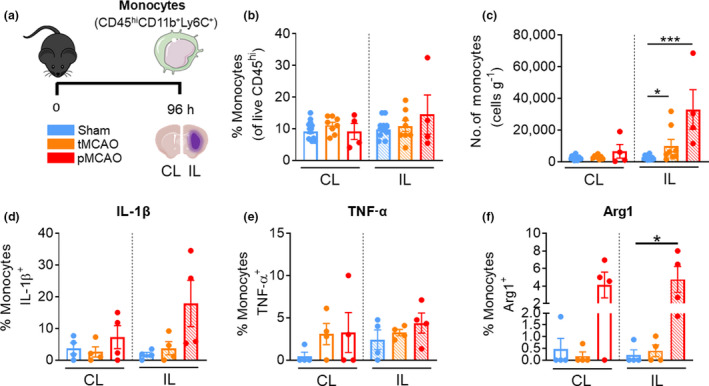
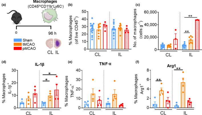
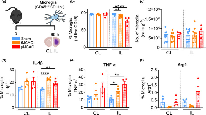
Previous studies investigating innate leukocyte recruitment into the brain after cerebral ischemia have shown conflicting results. Using distinct cell surface and intracellular markers, the current study evaluated the contributions of innate immune cells to the poststroke brain following 1-h middle cerebral artery occlusion (tMCAO) or permanent MCAO (pMCAO), and assessed whether these cells ascribed to an inflammatory state. Moreover, we examined whether there is evidence for leukocyte infiltration into the contralateral (CL) hemisphere despite the absence of stroke infarct. We observed the recruitment of peripheral neutrophils, monocytes and macrophages into the hemisphere ipsilateral (IL) to the ischemic brain infarct at 24 and 96 h following both tMCAO and pMCAO. In addition, we found evidence of increased leukocyte recruitment to the CL hemisphere but to a lesser extent than the IL hemisphere after stroke. Robust production of intracellular cytokines in the innate immune cell types examined was most evident at 24 h after pMCAO. Specifically, brain-associated neutrophils, monocytes and macrophages demonstrated stroke-induced production of tumor necrosis factor-α (TNF-α) and interleukin (IL)-1β, while only monocytes and macrophages exhibit a significant expression of arginase 1 (Arg1) after stroke. At 96 h after stroke, brain-resident microglia demonstrated production of TNF-α and IL-1β following both tMCAO and pMCAO. At this later timepoint, neutrophils displayed TNF-α production and brain-associated macrophages exhibited elevation of IL-1β and Arg1 after tMCAO. Further, pMCAO induced significant expression of Arg1 and IL-1β in monocytes and macrophages at 96 h, respectively. These results revealed that brain-associated innate immune cells display various stroke-induced inflammatory states that are dependent on the experimental stroke setting.
先前研究缺血性脑卒中后先天白细胞向脑内募集的结果存在争议。本研究使用不同的细胞表面和细胞内标志物,评估了 1 小时大脑中动脉闭塞(tMCAO)或永久性大脑中动脉闭塞(pMCAO)后卒中对大脑固有免疫细胞的影响,并评估这些细胞是否处于炎症状态。此外,我们还检查了尽管没有卒中梗死,但是否有白细胞浸润对侧(CL)半球的证据。我们观察到在 tMCAO 和 pMCAO 后 24 和 96 小时,缺血性脑梗死同侧(IL)半球有外周中性粒细胞、单核细胞和巨噬细胞募集。此外,我们发现卒中后 CL 半球的白细胞募集增加,但程度低于 IL 半球。在 pMCAO 后 24 小时,研究中检查的固有免疫细胞类型中细胞内细胞因子的产生明显增加。具体来说,与脑相关的中性粒细胞、单核细胞和巨噬细胞表现出卒中诱导的肿瘤坏死因子-α(TNF-α)和白细胞介素(IL)-1β的产生,而只有单核细胞和巨噬细胞在卒中后表现出显著的精氨酸酶 1(Arg1)表达。在卒中后 96 小时,脑驻留小胶质细胞在 tMCAO 和 pMCAO 后均表现出 TNF-α和 IL-1β的产生。在这个较晚的时间点,中性粒细胞表现出 TNF-α的产生,与脑相关的巨噬细胞在 tMCAO 后表现出 IL-1β和 Arg1 的升高。此外,pMCAO 在 96 小时分别诱导单核细胞和巨噬细胞中 Arg1 和 IL-1β的显著表达。这些结果表明,与脑相关的固有免疫细胞显示出各种依赖于实验性卒中设置的卒中诱导的炎症状态。