Department of Molecular, Cellular, and Developmental Biology, University of Michigan, 4104 Biological Sciences Building, 1105 North University Avenue, Ann Arbor, MI 48109, USA.
Department of Molecular and Human Genetics, Baylor College of Medicine, Jan and Dun Neurological Research Institute, Suite 1125, 1250 Mursund Street, Houston, TX 77030, USA.
STAR Protoc. 2022 Jun 10;3(2):101453. doi: 10.1016/j.xpro.2022.101453. eCollection 2022 Jun 17.
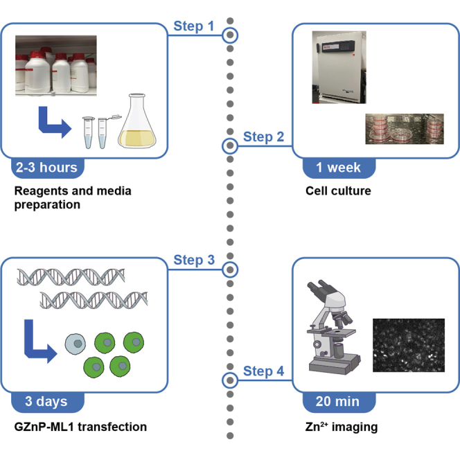
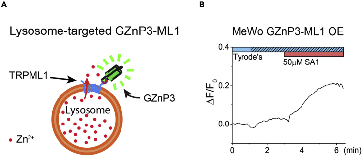
Intracellular vesicles such as lysosomes contain micromolar to millimolar concentrations of Zn, and disturbing lysosomal Zn homeostasis via lysosomal Zn release leads to mitochondria damage and consequent lytic cell death. Methods have been developed to image cellular Zn dynamics. Here, we present a protocol using GZnP3, a genetically encoded fluorescent Zn indicator, to assess lysosomal Zn release in cultured cells by fluorescence microscopy imaging. For complete details on the use and execution of this protocol, please refer to Du et al. (2021) or Minckley et al. (2019).
细胞内囊泡,如溶酶体,含有微摩尔到毫摩尔浓度的锌,通过溶酶体锌释放扰乱溶酶体锌稳态会导致线粒体损伤和随后的裂解性细胞死亡。已经开发出了用于成像细胞内锌动态变化的方法。在这里,我们通过荧光显微镜成像,使用 GZnP3(一种遗传编码的荧光锌指示剂)来介绍一种评估培养细胞中溶酶体锌释放的方案。有关该方案使用和执行的完整详细信息,请参考 Du 等人(2021 年)或 Minckley 等人(2019 年)。