Department of Oral Health Sciences and Department of Dentistry, Unit of Paediatric Dentistry and Special Dental Care, University Hospitals Leuven, KU Leuven, Kapucijnenvoer 7, PO box 7001, 3000, Leuven, Belgium.
OMFS IMPATH Research Group, Department of Imaging and Pathology, Faculty of Medicine, University of Leuven, Louvain, Belgium.
BMC Oral Health. 2022 Jun 20;22(1):245. doi: 10.1186/s12903-022-02281-4.
The aim of this study is to evaluate the impact of experience with traumatic dental injuries (TDI) on paediatric dentists' performance and self-assessed confidence when radiodiagnosing traumatic dental injuries (TDI) and to explore whether this is influenced by the imaging technique used (2D versus 3D).
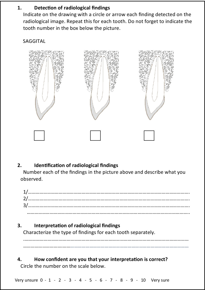
Both 2D and 3D radiological images of young anterior permanent teeth having experienced dental trauma were assessed randomly by a panel of paediatric dentists using structured scoring sheets. The impact of level of experience with dental traumatology on radiological detection, identification and interpretation of lesions and on observer's self-assessed confidence was evaluated. Findings were compared to benchmark data deriving from expert consensus of an experienced paediatric endodontologist and dentomaxillofacial radiologist. Results were analysed using generalized linear mixed modelling.
Overall, observers performed moderately to poor, irrespective of their level of TDI experience and imaging modality used. No proof could be yielded that paediatric dentists with high TDI experience performed better than those with low experience, for any of the outcomes and irrespective of the imaging modality used. When comparing the use of 3D images with 2D images, significantly higher sensitivities for the detection and correct identification of anomalies were observed in the low experienced group (P < 0.05). This was not the case regarding interpretation of the findings. Self-assessed confidence was significantly higher in more experienced dentists, both when using 2D and 3D images (P < 0.05).
There was no proof that paediatric dentist's higher experience with TDI is associated with better radiodiagnostic performance. Neither could it be proven that the use of Cone Beam Computed Tomography (CBCT) contributes to an improved interpretation of findings, for any experience level. More experienced dentists feel more confident, irrespective of the imaging modality used, but this does not correlate with improved performance. The overall poor performance in image interpretation highlights the importance of teaching and training in both dental radiology and dental traumatology.
本研究旨在评估创伤性牙外伤(TDI)经验对儿科牙医诊断创伤性牙外伤(TDI)的表现和自我评估信心的影响,并探讨使用的成像技术(二维与三维)是否对此有影响。
使用结构评分表,由一组儿科牙医对经历过牙外伤的年轻恒前牙的二维和三维放射影像学图像进行随机评估。评估创伤性牙外伤经验水平对病变的放射学检测、识别和解释以及观察者自我评估信心的影响。结果与经验丰富的儿科牙髓病学家和口腔颌面放射科医生的专家共识基准数据进行比较。结果使用广义线性混合模型进行分析。
总体而言,无论其 TDI 经验水平和使用的成像方式如何,观察者的表现均为中等至较差。对于任何结果,都没有证据表明高 TDI 经验的儿科牙医比低经验的牙医表现更好,也没有证据表明使用任何成像方式都如此。与二维图像相比,低经验组在检测和正确识别异常方面的敏感性明显更高(P<0.05),但在解释结果方面并非如此。自我评估的信心在经验更丰富的牙医中明显更高,无论是使用二维还是三维图像(P<0.05)。
没有证据表明儿科牙医更高的 TDI 经验与更好的放射诊断表现相关。也没有证据表明锥形束计算机断层扫描(CBCT)的使用有助于任何经验水平下对结果的解释。经验更丰富的牙医更有信心,无论使用何种成像方式,但这与表现的提高无关。总体上图像解释表现不佳突出了在牙科放射学和牙科创伤学方面进行教学和培训的重要性。