Department of Electrical Engineering, Columbia University, New York, NY, 10027, USA.
School of Integrated Circuits, Peking University, Beijing, P. R. China.
Nat Commun. 2022 Jun 20;13(1):3521. doi: 10.1038/s41467-022-31166-x.
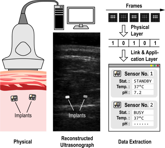
Modern clinical practice benefits significantly from imaging technologies and much effort is directed toward making this imaging more informative through the addition of contrast agents or reporters. Here, we report the design of a battery-less integrated circuit mote acting as an electronic reporter during medical ultrasound imaging. When implanted within the field-of-view of a brightness-mode (B-mode) ultrasound imager, this mote transmits information from its location through backscattered acoustic energy which is captured within the ultrasound image itself. We prototype and characterize the operation of such motes in vitro and in vivo. Performing with a static power consumption of less than 57 pW, the motes operate at duty cycles for receiving acoustic energy as low as 50 ppm. Motes within the same field-of-view during imaging have demonstrated signal-to-noise ratios of more than 19.1 dB at depths of up to 40 mm in lossy phantom. Physiological information acquired through such motes, which is beyond what is measurable with endogenous ultrasound backscatter and which is biogeographically located within an image, has the potential to provide an augmented ultrasonography.
现代临床实践显著受益于成像技术,人们投入大量精力通过添加造影剂或报告器使成像更具信息量。在这里,我们报告了一种无电池集成电路微电机的设计,该微电机在医学超声成象过程中充当电子报告器。当将此微电机植入亮度模式(B 模式)超声成像仪的视场范围内,它会通过反向散射声能将其位置信息传输到超声图像中。我们对这种微电机进行了原型设计和体内外特性分析。这种微电机的静态功耗小于 57pW,工作在接收声能的占空比低至 50ppm 的情况下。在成像过程中,同一视场范围内的微电机在 40mm 深的损耗性仿体中实现了超过 19.1dB 的信噪比。通过这种微电机获取的生理信息超出了内源性超声反向散射可测量的范围,并且在图像的生物地理定位范围内,有可能提供增强型超声成像。