Elliott Kiona, Berry Jeffrey C, Kim Hobin, Bart Rebecca S
Donald Danforth Plant Science Center, Saint Louis, MO, 63132, USA.
Division of Biological and Biomedical Sciences, Washington University in Saint Louis, St. Louis, MO, 63110, USA.
Plant Methods. 2022 Jun 21;18(1):86. doi: 10.1186/s13007-022-00906-x.
Methods to accurately quantify disease severity are fundamental to plant pathogen interaction studies. Commonly used methods include visual scoring of disease symptoms, tracking pathogen growth in planta over time, and various assays that detect plant defense responses. Several image-based methods for phenotyping of plant disease symptoms have also been developed. Each of these methods has different advantages and limitations which should be carefully considered when choosing an approach and interpreting the results.
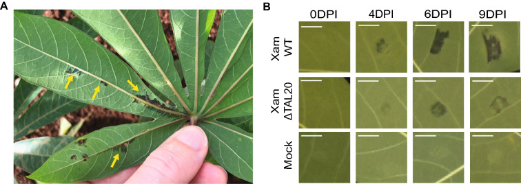
In this paper, we developed two image analysis methods and tested their ability to quantify different aspects of disease lesions in the cassava-Xanthomonas pathosystem. The first method uses ImageJ, an open-source platform widely used in the biological sciences. The second method is a few-shot support vector machine learning tool that uses a classifier file trained with five representative infected leaf images for lesion recognition. Cassava leaves were syringe infiltrated with wildtype Xanthomonas, a Xanthomonas mutant with decreased virulence, and mock treatments. Digital images of infected leaves were captured overtime using a Raspberry Pi camera. The image analysis methods were analyzed and compared for the ability to segment the lesion from the background and accurately capture and measure differences between the treatment types.
Both image analysis methods presented in this paper allow for accurate segmentation of disease lesions from the non-infected plant. Specifically, at 4-, 6-, and 9-days post inoculation (DPI), both methods provided quantitative differences in disease symptoms between different treatment types. Thus, either method could be applied to extract information about disease severity. Strengths and weaknesses of each approach are discussed.
准确量化疾病严重程度的方法是植物病原体相互作用研究的基础。常用方法包括对疾病症状进行视觉评分、随时间跟踪植物体内病原体的生长,以及检测植物防御反应的各种测定方法。还开发了几种基于图像的植物病害症状表型分析方法。这些方法各有不同的优点和局限性,在选择方法和解释结果时应仔细考虑。
在本文中,我们开发了两种图像分析方法,并测试了它们量化木薯 - 黄单胞菌病理系统中病害病变不同方面的能力。第一种方法使用ImageJ,这是一个在生物科学中广泛使用的开源平台。第二种方法是一种少样本支持向量机学习工具,它使用一个用五张代表性感染叶片图像训练的分类器文件进行病变识别。用野生型黄单胞菌、毒力降低的黄单胞菌突变体和模拟处理对木薯叶片进行注射器浸润。使用树莓派相机随时间捕获感染叶片的数字图像。对图像分析方法进行了分析和比较,以评估其从背景中分割病变以及准确捕获和测量不同处理类型之间差异的能力。
本文提出的两种图像分析方法都能准确地从未感染植物中分割出病害病变。具体而言,在接种后4天、6天和9天,两种方法都提供了不同处理类型之间疾病症状的定量差异。因此,这两种方法都可用于提取有关疾病严重程度的信息。讨论了每种方法的优缺点。