Department of Obstetrics and Gynecology, School of Medicine, Shimane University, 89-1 Enya Cho, Izumo, Shimane, 693-8501, Japan.
Reprod Biol Endocrinol. 2022 Jun 21;20(1):91. doi: 10.1186/s12958-022-00963-w.
Kisspeptin released from Kiss-1 neurons in the hypothalamus plays an essential role in the control of the hypothalamic-pituitary-gonadal axis by regulating the release of gonadotropin-releasing hormone (GnRH). In this study, we examined how androgen supplementation affects the characteristics of Kiss-1 neurons.
We used a Kiss-1-expressing mHypoA-55 cell model that originated from the arcuate nucleus (ARC) of the mouse hypothalamus. These cells are KNDy neurons that co-express neurokinin B (NKB) and dynorphin A (DynA). We stimulated these cells with androgens and examined them. We also examined the ARC region of the hypothalamus in ovary-intact female rats after supplementation with androgens.
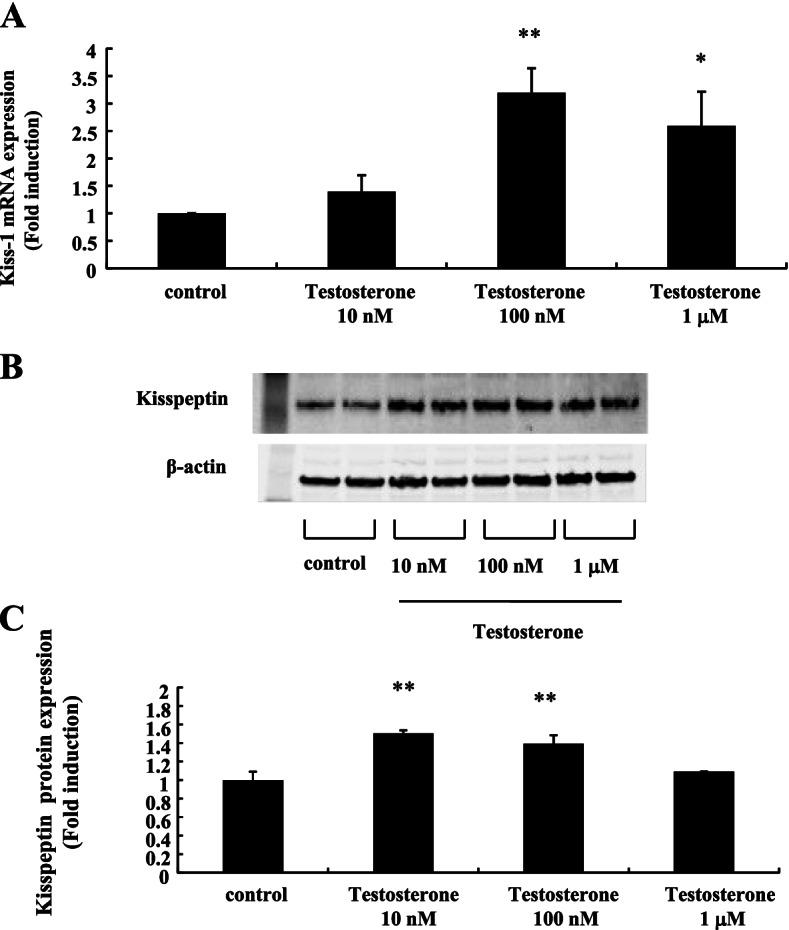
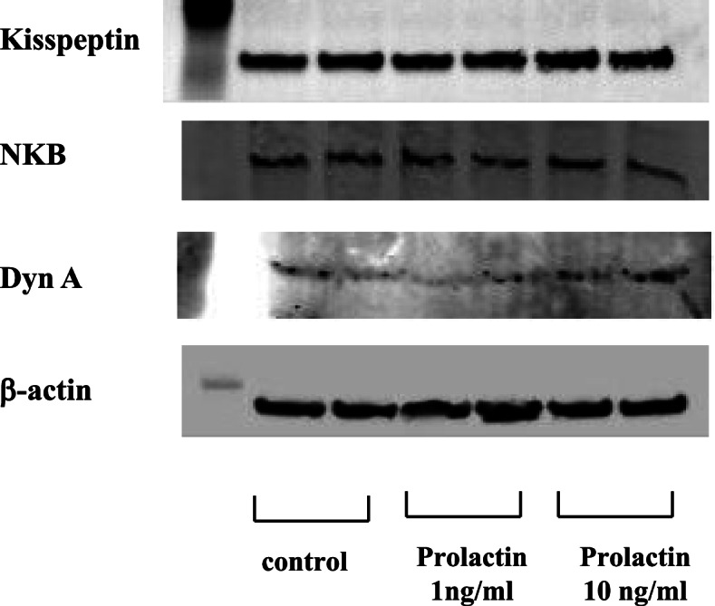
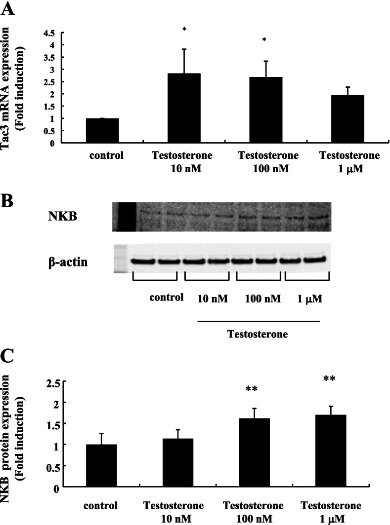
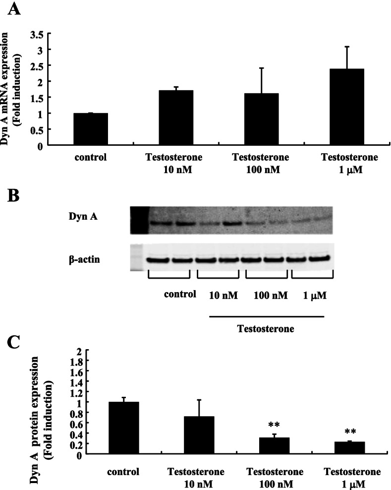
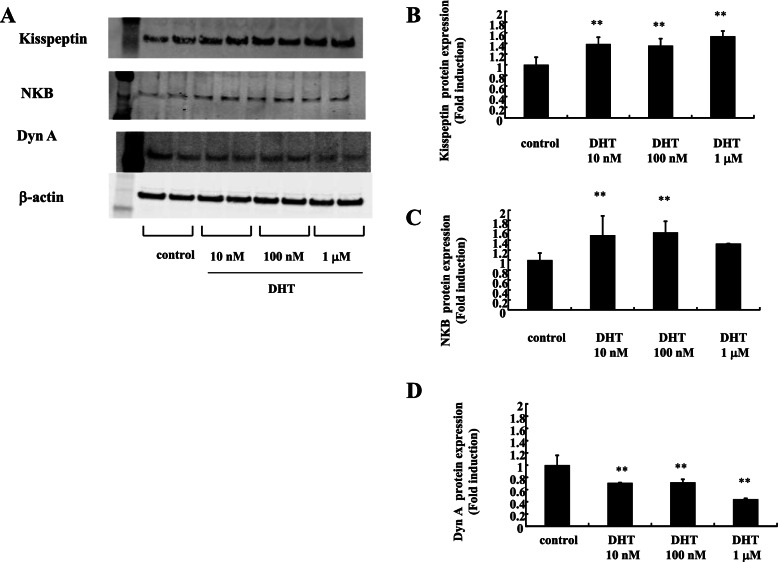
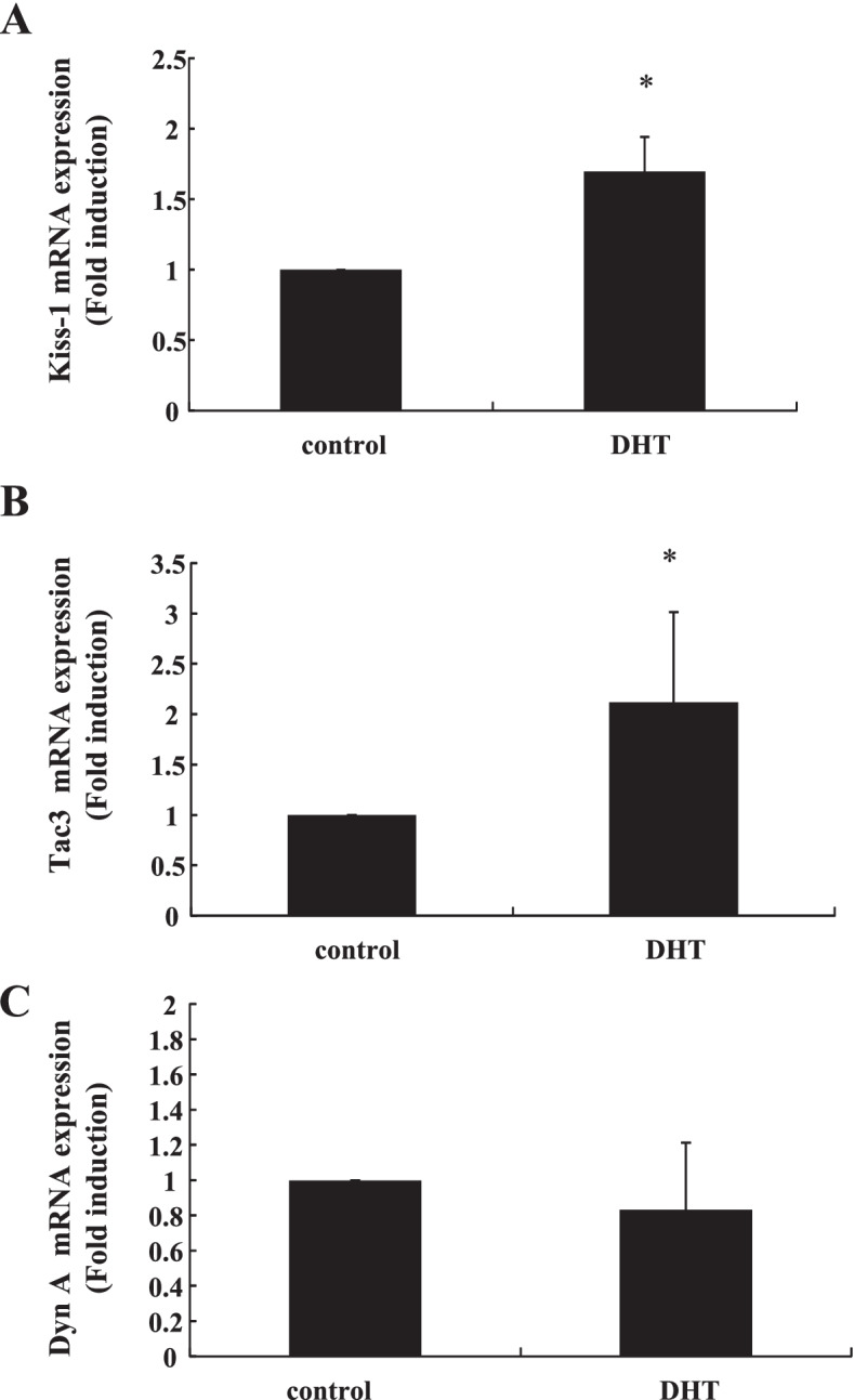
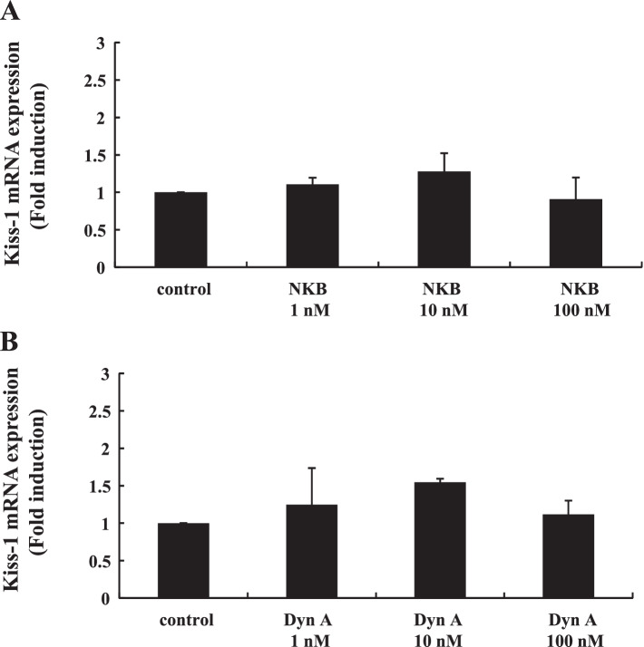
Stimulation of mHypoA-55 cells with 100 nM testosterone significantly increased Kiss-1 gene expression by 3.20 ± 0.44-fold; testosterone also increased kisspeptin protein expression. The expression of Tac3, the gene encoding NKB, was also increased by 2.69 ± 0.64-fold following stimulation of mHypoA-55 cells with 100 nM testosterone. DynA gene expression in these cells was unchanged by testosterone stimulation, but it was significantly reduced at the protein level. Dihydrotestosterone (DHT) had a similar effect to testosterone in mHypoA-55 cells; kisspeptin and NKB protein expression was significantly increased by DHT, whereas it significantly reduced DynA expression. In ovary-intact female rats, DTH administration significantly increased the gene expression of Kiss-1 and Tac3, but not DynA, in the arcuate nucleus. Exogenous NKB and DynA stimulation failed to modulate Kiss-1 gene expression in mHypoA-55 cells. Unlike androgen stimulation, prolactin stimulation did not modulate kisspeptin, NKB, or DynA protein expression in these cells.
Our observations imply that hyperandrogenemia affects KNDy neurons and changes their neuronal characteristics by increasing kisspeptin and NKB levels and decreasing DynA levels. These changes might cause dysfunction of the hypothalamic-pituitary-gonadal axis.
下丘脑 Kiss-1 神经元释放的 Kisspeptin 通过调节促性腺激素释放激素 (GnRH) 的释放,在控制下丘脑-垂体-性腺轴中发挥着重要作用。在这项研究中,我们研究了雄激素补充如何影响 Kiss-1 神经元的特征。
我们使用了一种源于小鼠下丘脑弓状核 (ARC) 的表达 Kiss-1 的 mHypoA-55 细胞模型。这些细胞是共表达神经激肽 B (NKB) 和强啡肽 A (DynA) 的 KNDy 神经元。我们用雄激素刺激这些细胞并进行了检查。我们还检查了卵巢完整的雌性大鼠补充雄激素后下丘脑 ARC 区域的情况。
100 nM 睾酮刺激 mHypoA-55 细胞可使 Kiss-1 基因表达显著增加 3.20±0.44 倍;睾酮还增加了 kisspeptin 蛋白表达。100 nM 睾酮刺激 mHypoA-55 细胞后,Tac3(编码 NKB 的基因)的表达也增加了 2.69±0.64 倍。这些细胞中 DynA 基因的表达不受睾酮刺激的影响,但在蛋白质水平上显著降低。二氢睾酮 (DHT) 在 mHypoA-55 细胞中也具有类似的作用;DHT 显著增加 kisspeptin 和 NKB 蛋白表达,而显著降低 DynA 表达。在卵巢完整的雌性大鼠中,DTH 给药显著增加了 ARC 中 Kiss-1 和 Tac3 的基因表达,但 DynA 没有增加。外源性 NKB 和 DynA 刺激未能调节 mHypoA-55 细胞中 Kiss-1 的基因表达。与雄激素刺激不同,催乳素刺激不能调节这些细胞中的 kisspeptin、NKB 或 DynA 蛋白表达。
我们的观察结果表明,高雄激素血症通过增加 kisspeptin 和 NKB 水平以及降低 DynA 水平影响 KNDy 神经元并改变其神经元特征。这些变化可能导致下丘脑-垂体-性腺轴功能障碍。