Department of Radiology and Biomedical Imaging, University of California, 185 Berry Street, San Francisco, CA, 94107, USA.
Department of Nuclear Engineering, University of California, Berkeley, CA, USA.
Eur J Nucl Med Mol Imaging. 2022 Sep;49(11):3761-3771. doi: 10.1007/s00259-022-05858-x. Epub 2022 Jun 23.
Non-invasive imaging is a key clinical tool for detection and treatment monitoring of infections. Existing clinical imaging techniques are frequently unable to distinguish infection from tumors or sterile inflammation. This challenge is well-illustrated by prosthetic joint infections that often complicate joint replacements. D-methyl-C-methionine (D-C-Met) is a new bacteria-specific PET radiotracer, based on an amino acid D-enantiomer, that is rapidly incorporated into the bacterial cell wall. In this manuscript, we describe the biodistribution, radiation dosimetry, and initial human experience using D-C-Met in patients with suspected prosthetic joint infections.
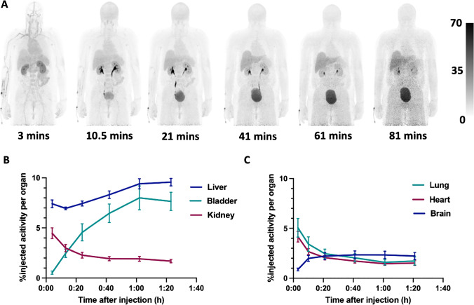
614.5 ± 100.2 MBq of D-C-Met was synthesized using an automated in-loop radiosynthesis method and administered to six healthy volunteers and five patients with suspected prosthetic joint infection, who were studied by PET/MRI. Time-activity curves were used to calculate residence times for each source organ. Absorbed doses to each organ and body effective doses were calculated using OLINDA/EXM 1.1 with both ICRP 60 and ICRP 103 tissue weighting factors. SUV and SUV were calculated for volumes of interest (VOIs) in joints with suspected infection, the unaffected contralateral joint, blood pool, and soft tissue background. A two-tissue compartment model was used for kinetic modeling.
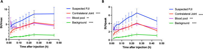
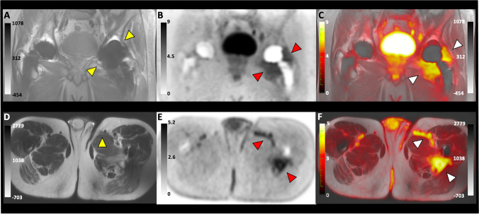
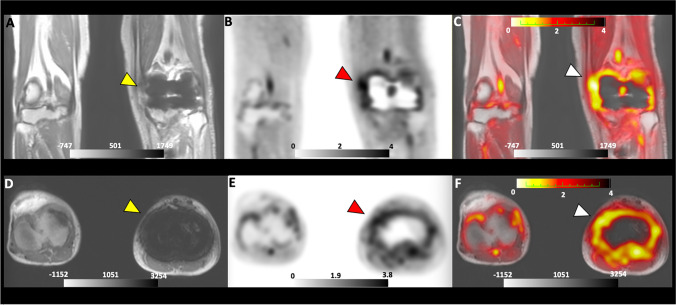
D-C-Met was well tolerated in all subjects. The tracer showed clearance from both urinary (rapid) and hepatobiliary (slow) pathways as well as low effective doses. Moreover, minimal background was observed in both organs with resident micro-flora and target organs, such as the spine and musculoskeletal system. Additionally, D-C-Met showed increased focal uptake in areas of suspected infection, demonstrated by a significantly higher SUV and SUV calculated from VOIs of joints with suspected infections compared to the contralateral joints, blood pool, and background (P < 0.01). Furthermore, higher distribution volume and binding potential were observed in suspected infections compared to the unaffected joints.
D-C-Met has a favorable radiation profile, minimal background uptake, and fast urinary extraction. Furthermore, D-C-Met showed increased uptake in areas of suspected infection, making this a promising approach. Validation in larger clinical trials with a rigorous gold standard is still required.
非侵入性成像技术是检测和治疗感染的重要临床工具。现有的临床成像技术常常无法区分感染、肿瘤或无菌性炎症。这一挑战在人工关节感染中得到了很好的体现,这种感染常导致关节置换术后并发症。D-甲基-C-蛋氨酸(D-C-Met)是一种新的细菌特异性 PET 放射性示踪剂,基于氨基酸的 D-对映体,可迅速掺入细菌细胞壁。在本文中,我们描述了 D-C-Met 的生物分布、辐射剂量学以及在疑似人工关节感染患者中的初步人体经验。
使用自动环内放射合成方法合成了 614.5±100.2 MBq 的 D-C-Met,并在 6 名健康志愿者和 5 名疑似人工关节感染的患者中进行了 PET/MRI 研究。使用时间-活性曲线计算每个源器官的居留时间。使用 OLINDA/EXM 1.1 计算每个器官的吸收剂量和体有效剂量,分别采用 ICRP 60 和 ICRP 103 组织权重因子。对可疑感染关节、未受影响的对侧关节、血池和软组织背景的感兴趣体积(VOI)计算 SUV 和 SUV。使用双组织室模型进行动力学建模。
D-C-Met 在所有受试者中均耐受良好。该示踪剂从尿(快速)和肝胆(缓慢)途径清除,有效剂量低。此外,在具有常驻微生物群和目标器官(如脊柱和肌肉骨骼系统)的器官中,观察到的背景最小。此外,在疑似感染区域,D-C-Met 显示出明显的局灶性摄取增加,这可通过对疑似感染关节的 VOI 与对侧关节、血池和背景相比计算出的 SUV 和 SUV 显著升高来证明(P<0.01)。此外,与未受影响的关节相比,在疑似感染部位观察到更高的分布容积和结合潜能。
D-C-Met 具有良好的辐射特性、最小的背景摄取和快速的尿液提取。此外,D-C-Met 在疑似感染区域的摄取增加,这是一种很有前途的方法。仍需要在严格的金标准的更大临床试验中进行验证。