Division of Infectious Diseases, Department of Pediatrics, Johns Hopkins University School of Medicine, Baltimore, Maryland, USA.
Center for Infection and Inflammation Imaging Research, Johns Hopkins University School of Medicine, Baltimore, Maryland, USA.
mBio. 2019 Oct 29;10(5):e00317-19. doi: 10.1128/mBio.00317-19.
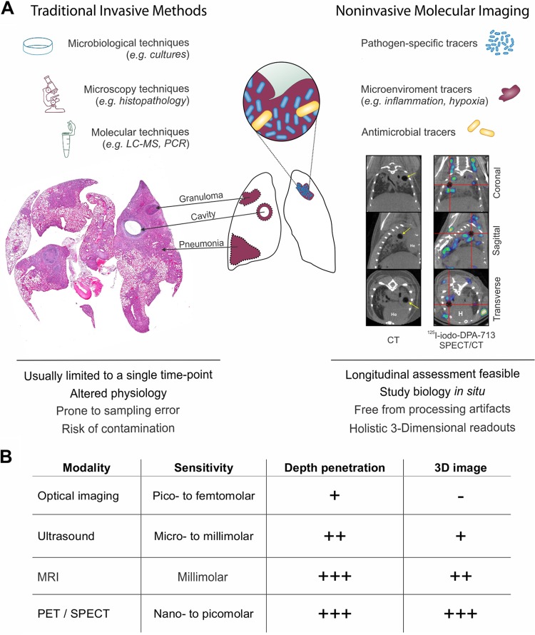
Molecular imaging is an emerging technology that enables the noninvasive visualization, characterization, and quantification of molecular events within living subjects. Positron emission tomography (PET) is a clinically available molecular imaging tool with significant potential to study pathogenesis of infections in humans. PET enables dynamic assessment of infectious processes within the same subject with high temporal and spatial resolution and obviates the need for invasive tissue sampling, which is difficult in patients and generally limited to a single time point, even in animal models. This review presents current state-of-the-art concepts on the application of molecular imaging for infectious diseases and details how PET imaging can facilitate novel insights into infectious processes, ongoing development of pathogen-specific imaging, and simultaneous measurements of intralesional antimicrobial pharmacokinetics in multiple compartments, including privileged sites. Finally, the potential clinical applications of this promising technology are also discussed.
分子成像技术是一种新兴的技术,可实现活体生物体内分子事件的非侵入性可视化、特征描述和定量分析。正电子发射断层扫描(PET)是一种临床可用的分子成像工具,具有研究人类感染发病机制的巨大潜力。PET 能够以高时间和空间分辨率动态评估同一患者体内的感染过程,并且避免了对难以在患者中进行的侵入性组织采样的需求,而这种采样通常限于单个时间点,即使在动物模型中也是如此。本综述介绍了分子成像在传染病中的最新应用概念,并详细说明了 PET 成像如何有助于深入了解感染过程、正在开发的针对病原体的成像方法,以及同时在多个隔室(包括特权部位)中测量病灶内抗菌药代动力学的新方法。最后,还讨论了这项有前途的技术的潜在临床应用。