Helmedag Marius J, Heise Daniel, Eickhoff Roman M, Schmitz Sophia M, Mechelinck Mare, Emonts Caroline, Bolle Tim, Gries Thomas, Neumann Ulf Peter, Klink Christian Daniel, Lambertz Andreas
Department of General, Visceral and Transplantation Surgery, RWTH Aachen University Hospital, 52074 Aachen, Germany.
Department of Anesthesiology, Uniklinik RWTH Aachen, 52074 Aachen, Germany.
Biomedicines. 2022 May 31;10(6):1294. doi: 10.3390/biomedicines10061294.
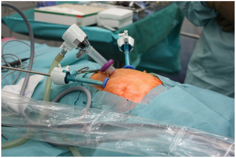
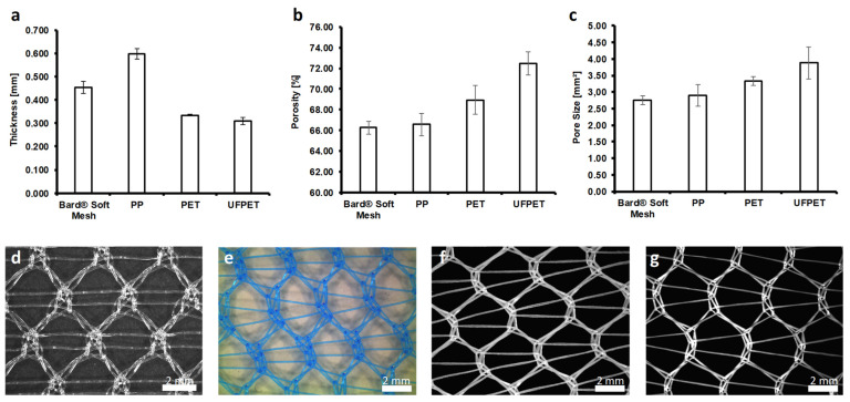
(1) Introduction: The intraperitoneal onlay mesh technique (IPOM) is widely used to repair incisional hernias. This method has advantages but suffers from complications due to intraperitoneal adhesion formation between the mesh and intestine. An ideal mesh minimizes adhesions and shows good biocompatibility. To address this, newly developed multifilamentous polyethylene (PET) meshes were constructed from sub-macrophage-sized monofilaments and studied regarding biocompatibility and adhesion formation. (2) Methods: We investigated fine (FPET, 72 filaments, 11 µm diameter each) and ultra-fine multifilament (UFPET, 700 filaments, 3 µm diameter each) polyethylene meshes for biocompatibility in subcutaneous implantation in rats. Adhesion formation was analyzed in the IPOM position in rabbits. Geometrically identical mono-filamentous polypropylene (PP) Bard Soft PP meshes were used for comparison. Histologic and immune-histologic foreign body reactions were assessed in 48 rats after 7 or 21 days (four mesh types, with two different mesh types per rat; = 6 per mesh type). Additionally, two different mesh types each were placed in the IPOM position in 24 rabbits to compile the Diamond peritoneal adhesion score after the same timeframes. The biocompatibility and adhesion score differences were analyzed with the Kruskal-Wallis nonparametric statistical test. (3) Results: Overall, FPET and, especially, UFPET showed significantly smaller foreign body granulomas compared to PP meshes. Longer observation periods enhanced the differences. Immunohistology showed no significant differences in the cellular immune response and proliferation. UFPET demonstrated significantly reduced peritoneal adhesion formation compared to all other tested meshes after 21 days. (4) Conclusions: Overall, FPET and, especially, UFPET demonstrated their suitability for IPOM hernia meshes in animal models by improving major aspects of the foreign body reaction and reducing adhesion formation.
(1) 引言:腹腔内置网技术(IPOM)广泛用于修补切口疝。该方法具有优点,但因网片与肠管之间形成腹腔内粘连而存在并发症。理想的网片可使粘连最小化并具有良好的生物相容性。为解决这一问题,用亚巨噬细胞大小的单丝构建了新开发的多丝状聚乙烯(PET)网片,并对其生物相容性和粘连形成进行了研究。(2) 方法:我们研究了细(FPET,72 根丝,每根直径 11 µm)和超细多丝(UFPET,700 根丝,每根直径 3 µm)聚乙烯网片在大鼠皮下植入中的生物相容性。在兔的 IPOM 位置分析粘连形成情况。使用几何形状相同的单丝状聚丙烯(PP)巴德软质 PP 网片作对照。在 7 天或 21 天后对 48 只大鼠(四种网片类型,每只大鼠使用两种不同网片类型;每种网片类型 = 6 只)的组织学和免疫组织学异物反应进行评估。此外,将每种两种不同网片类型置于 24 只兔的 IPOM 位置,在相同时间段后编制钻石腹膜粘连评分。用 Kruskal-Wallis 非参数统计检验分析生物相容性和粘连评分差异。(3) 结果:总体而言,与 PP 网片相比,FPET 尤其是 UFPET 显示出明显更小的异物肉芽肿。更长的观察期使差异增大。免疫组织学显示细胞免疫反应和增殖无显著差异。与所有其他测试网片相比,21 天后 UFPET 显示腹膜粘连形成显著减少。(