Li Yong, Zheng Feihui, Foo Li Lian, Wong Qiu Ying, Ting Daniel, Hoang Quan V, Chong Rachel, Ang Marcus, Wong Chee Wai
Singapore National Eye Centre, Singapore Eye Research Institute, Singapore 169856, Singapore.
Ophthalmology and Visual Sciences Academic Clinical Program, Duke-NUS Medical School, Singapore 169857, Singapore.
Diagnostics (Basel). 2022 Jun 8;12(6):1418. doi: 10.3390/diagnostics12061418.
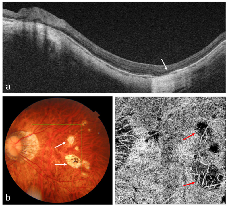
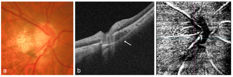
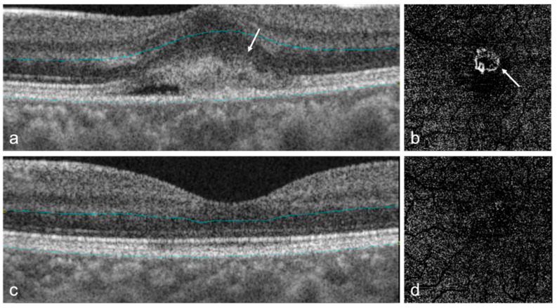
Advances in imaging with optical coherence tomography (OCT) and optical coherence tomography angiography (OCTA) technology, including the development of swept source OCT/OCTA, widefield or ultra-widefield systems, have greatly improved the understanding, diagnosis, and treatment of myopia and myopia-related complications. Anterior segment OCT is useful for imaging the anterior segment of myopes, providing the basis for implantable collamer lens optimization, or detecting intraocular lens decentration in high myopic patients. OCT has enhanced imaging of vitreous properties, and measurement of choroidal thickness in myopic eyes. Widefield OCT systems have greatly improved the visualization of peripheral retinal lesions and have enabled the evaluation of wide staphyloma and ocular curvature. Based on OCT imaging, a new classification system and guidelines for the management of myopic traction maculopathy have been proposed; different dome-shaped macula morphologies have been described; and myopia-related abnormalities in the optic nerve and peripapillary region have been demonstrated. OCTA can quantitatively evaluate the retinal microvasculature and choriocapillaris, which is useful for the early detection of myopic choroidal neovascularization and the evaluation of anti-vascular endothelial growth factor therapy in these patients. In addition, the application of artificial intelligence in OCT/OCTA imaging in myopia has achieved promising results.
光学相干断层扫描(OCT)和光学相干断层扫描血管造影(OCTA)技术的成像进展,包括扫频源OCT/OCTA、广角或超广角系统的发展,极大地提高了对近视及近视相关并发症的认识、诊断和治疗水平。眼前段OCT有助于对近视患者的眼前段进行成像,为可植入角膜接触镜的优化提供依据,或检测高度近视患者的人工晶状体偏心情况。OCT增强了对玻璃体特性的成像以及对近视眼脉络膜厚度的测量。广角OCT系统极大地改善了周边视网膜病变的可视化,并能够评估宽视盘和眼曲率。基于OCT成像,已经提出了一种新的近视性牵引性黄斑病变分类系统和管理指南;描述了不同的穹窿状黄斑形态;并展示了视神经和视乳头周围区域与近视相关的异常情况。OCTA可以定量评估视网膜微血管和脉络膜毛细血管,这有助于早期检测近视性脉络膜新生血管,并评估这些患者的抗血管内皮生长因子治疗效果。此外,人工智能在近视的OCT/OCTA成像中的应用已经取得了有前景的成果。