Castagneto-Gissey Lidia, Russo Maria Francesca, Iodice Alessandra, Casella-Mariolo James, Serao Angelo, Picchetto Andrea, D'Ambrosio Giancarlo, Urciuoli Irene, De Luca Alessandro, Salvati Bruno, Casella Giovanni
Department of Surgical Sciences, Sapienza University of Rome, 00161 Rome, Italy.
Department of General and Emergency Surgery, Ospedale dei Castelli (NOC), ASL Roma 6, 00040 Rome, Italy.
J Clin Med. 2022 Jun 17;11(12):3508. doi: 10.3390/jcm11123508.
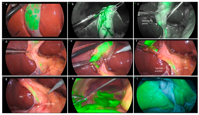
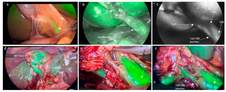
(1) Background: Fluorescence cholangiography has been proposed as a method for improving the visualization and identification of extrahepatic biliary anatomy in order to possibly reduce injuries and related complications. The most common method of indocyanine green (ICG) administration is the intravenous route, whereas evidence on direct ICG injection into the gallbladder is still quite limited. We aimed to compare the two different methods of ICG administration in terms of the visualization of extrahepatic biliary anatomy during laparoscopic cholecystectomy (LC), analyzing differences in the time of visualization, as well as the efficacy, advantages, and disadvantages of both modalities. (2) Methods: A total of 35 consecutive adult patients affected by acute or chronic gallbladder disease were enrolled in this prospective case−control study. Seventeen patients underwent LC with direct gallbladder ICG injection (IC-ICG) and eighteen subjects received intravenous ICG administration (IV-ICG). (3) Results: The groups were comparable with regard to their demographic and perioperative characteristics. The IV-ICG group had a significantly shorter overall operative time compared to the IC-ICG group (p = 0.017). IV-ICG was better at delineating the duodenum and the common hepatic duct compared to the IC-ICG method (p = 0.009 and p = 0.041, respectively). The cystic duct could be delineated pre-dissection in 76.5% and 66.7% of cases in the IC-ICG and IV-ICG group, respectively, and this increased to 88.2% and 83.3% after dissection. The common bile duct could be highlighted in 76.5% and 77.8% of cases in the IC-ICG and IV-ICG group, respectively. Liver fluorescence was present in one case in the IC-ICG group and in all cases after IV-ICG administration (5.8% versus 100%; p < 0.0001). (4) Conclusions: The present study demonstrates how ICG-fluorescence cholangiography can be helpful in identifying the extrahepatic biliary anatomy during dissection of Calot’s triangle in both administration methods. In comparison with intravenous ICG injection, the intracholecystic ICG route could provide a better signal-to-background ratio by avoiding hepatic fluorescence, thus increasing the bile duct-to-liver contrast.
(1)背景:荧光胆管造影术已被提议作为一种改善肝外胆管解剖结构可视化和识别的方法,以期减少损伤及相关并发症。吲哚菁绿(ICG)给药的最常见方法是静脉途径,而关于直接将ICG注入胆囊的证据仍然相当有限。我们旨在比较两种不同的ICG给药方法在腹腔镜胆囊切除术(LC)期间肝外胆管解剖结构可视化方面的差异,分析可视化时间的差异以及两种方式的疗效、优点和缺点。(2)方法:本前瞻性病例对照研究共纳入35例连续的患有急性或慢性胆囊疾病的成年患者。17例患者接受了直接胆囊内注入ICG的LC手术(IC-ICG组),18例患者接受了静脉注射ICG(IV-ICG组)。(3)结果:两组在人口统计学和围手术期特征方面具有可比性。与IC-ICG组相比,IV-ICG组的总手术时间明显更短(p = 0.017)。与IC-ICG方法相比,IV-ICG在勾勒十二指肠和肝总管方面表现更好(分别为p = 0.009和p = 0.041)。在IC-ICG组和IV-ICG组中,分别有76.5%和66.7%的病例在胆囊管解剖前可勾勒出胆囊管,解剖后这一比例分别增至88.2%和83.3%。在IC-ICG组和IV-ICG组中,分别有76.5%和77.8%的病例可突出显示胆总管。IC-ICG组有1例出现肝脏荧光,IV-ICG给药后所有病例均出现肝脏荧光(5.8%对100%;p < 0.0001)。(4)结论:本研究表明,在两种给药方法中,ICG荧光胆管造影术在解剖Calot三角期间识别肝外胆管解剖结构方面均有帮助。与静脉注射ICG相比,胆囊内ICG途径可通过避免肝脏荧光提供更好的信号背景比,从而增加胆管与肝脏的对比度。