Andresciani Flavio, Ricci Milena, Grasso Rosario Francesco, Zobel Bruno Beomonte, Quattrocchi Carlo Cosimo
Unit of Diagnostic Imaging and Interventional Radiology, Fondazione Policlinico Campus Bio-Medico di Roma, Università Campus Bio-Medico di Roma, via Alvaro del Portillo 21, Rome 00128, Italy.
Radiol Case Rep. 2022 Jun 17;17(9):2996-2999. doi: 10.1016/j.radcr.2022.05.072. eCollection 2022 Sep.
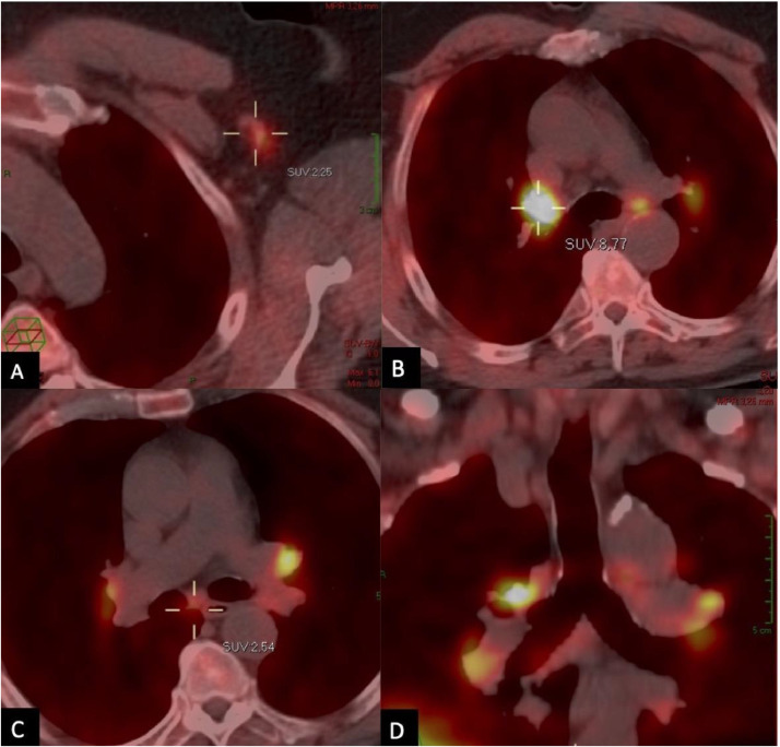
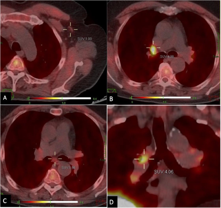
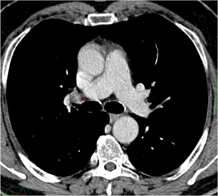
Several cases of cancer patients with 18-fluorodeoxyglucose (FDG) Positron Emission Tomography/Computed Tomography (PET/CT) evidence of metabolically active axillary lymph nodes after COVID-19 vaccination have been described, creating a diagnostic dilemma and sometimes leading to further unnecessary examinations. A 62-year-old male, diagnosed with prostate cancer, treated with hormone-therapy and radiotherapy of the prostate 2 years before, underwent fluorine-18 choline (F-FCH) PET/CT for restaging purpose, less than 3 weeks after he had received the second dose of the Pfizer BioNTech-BNT162b2 mRNA COVID-19 vaccine. This exam showed an increased F-FCH uptake and an enlargement of the left axillary, paratracheal, para-aortic, subcarinal, and hilar bilateral lymph nodes. Fourteen weeks later, the patient underwent a new F-FCH PET-CT scan, displaying an almost complete regularization of the FCH uptake in all the previously involved regions. The patient was not treated after the first PET-CT scan, thus, the aforementioned PET/CT findings represented inflammatory vaccine-related lymph nodes. This case highlights the significance of knowing vaccination history to correctly interpret imaging findings and to avoid false-positive reports.
已有多例癌症患者在接种新冠病毒疫苗后,18-氟脱氧葡萄糖(FDG)正电子发射断层扫描/计算机断层扫描(PET/CT)显示腋窝淋巴结代谢活跃,这造成了诊断困境,有时还会导致进一步的不必要检查。一名62岁男性,2年前被诊断为前列腺癌,接受了前列腺激素治疗和放疗,在接种辉瑞BioNTech-BNT162b2 mRNA新冠病毒疫苗第二剂后不到3周,为进行再分期接受了氟-18胆碱(F-FCH)PET/CT检查。该检查显示F-FCH摄取增加,左侧腋窝、气管旁、主动脉旁、隆突下及双侧肺门淋巴结肿大。14周后,患者接受了新的F-FCH PET-CT扫描,显示所有先前受累区域的FCH摄取几乎完全恢复正常。首次PET-CT扫描后患者未接受治疗,因此,上述PET/CT表现代表与疫苗相关的炎性淋巴结。该病例强调了了解疫苗接种史对于正确解读影像学结果和避免假阳性报告的重要性。