College of Computer Science and Technology, Zhejiang University of Technology, Hangzhou, China.
J Appl Clin Med Phys. 2022 Aug;23(8):e13709. doi: 10.1002/acm2.13709. Epub 2022 Jun 24.
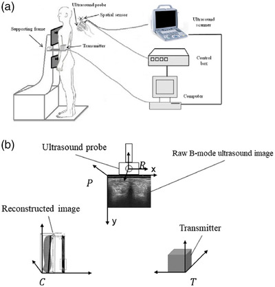
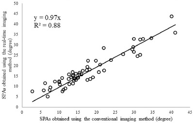
Real-time 3D ultrasound has gained popularity in many fields because it can provide interactive feedback to help acquire high-quality images or to conduct timely diagnosis. However, no comprehensive study has been reported on such an imaging method for scoliosis evaluation due to the complexity of this application. Meanwhile, the use of radiation-free assessment of scoliosis is becoming increasingly popular. This study developed a real-time 3D ultrasound imaging method for scoliosis assessment based on an incremental imaging method. In vivo experiments involving 36 patients with scoliosis were performed to test the performance of the proposed method. This new imaging method achieved a mean incremental frame rate of 82.7 ± 11.0 frames/s. The high repeatability of the intra-operator test (intraclass correlation coefficient [ICC] = 0.92) and inter-operator test (ICC = 0.91) demonstrated that the new method was very reliable. The result of spinous process angles obtained by the new method was linearly correlated (y = 0.97x, R = 0.88) with that obtained by conventional 3D reconstruction. These results suggested that the newly developed imaging method can provide real-time ultrasound imaging for scoliosis evaluation while preserving the comparative image quality of the conventional 3D reconstruction method.
实时 3D 超声在许多领域中得到了广泛应用,因为它可以提供交互式反馈,帮助获取高质量的图像或进行及时的诊断。然而,由于这种应用的复杂性,目前还没有关于这种成像方法用于脊柱侧凸评估的全面研究。同时,无辐射评估脊柱侧凸的方法也越来越受到关注。本研究基于增量成像方法开发了一种用于脊柱侧凸评估的实时 3D 超声成像方法。对 36 例脊柱侧凸患者进行了体内实验,以测试所提出方法的性能。该新的成像方法实现了 82.7±11.0 帧/秒的平均增量帧率。内-操作者测试(组内相关系数 [ICC]=0.92)和间-操作者测试(ICC=0.91)的高重复性表明该新方法非常可靠。新方法获得的棘突角度的结果与传统的 3D 重建方法呈线性相关(y=0.97x,R=0.88)。这些结果表明,新开发的成像方法可以为脊柱侧凸评估提供实时超声成像,同时保持传统 3D 重建方法的可比图像质量。