• 文献检索
  • 文档翻译
  • 深度研究
  • 学术资讯
  • Suppr Zotero 插件Zotero 插件
  • 邀请有礼
  • 套餐&价格
  • 历史记录
应用&插件
Suppr Zotero 插件Zotero 插件浏览器插件Mac 客户端Windows 客户端微信小程序
定价
高级版会员购买积分包购买API积分包
服务
文献检索文档翻译深度研究API 文档MCP 服务
关于我们
关于 Suppr公司介绍联系我们用户协议隐私条款
关注我们

Suppr 超能文献

核心技术专利:CN118964589B侵权必究
粤ICP备2023148730 号-1Suppr @ 2026

文献检索

告别复杂PubMed语法,用中文像聊天一样搜索,搜遍4000万医学文献。AI智能推荐,让科研检索更轻松。

立即免费搜索

文件翻译

保留排版,准确专业,支持PDF/Word/PPT等文件格式,支持 12+语言互译。

免费翻译文档

深度研究

AI帮你快速写综述,25分钟生成高质量综述,智能提取关键信息,辅助科研写作。

立即免费体验

白内障手术中三维手术系统与双目显微镜用于透明角膜切口的比较。

Comparison of Three-Dimensional Surgical System Versus Binocular Microscope for Clear Corneal Incision in Cataract Surgery.

作者信息

Zhu Zehui, Chang Pingjun, Huang Feng, Shen Songqing, Zhao Xiaomeng, Ji Xinpei, Zhao Yun E

机构信息

Eye Hospital and School of Ophthalmology and Optometry, Wenzhou Medical University, Wenzhou, Zhejiang, China.

National Clinical Research Center for Ocular Diseases, Wenzhou, Zhejiang, China.

出版信息

Ophthalmol Ther. 2022 Aug;11(4):1589-1600. doi: 10.1007/s40123-022-00537-4. Epub 2022 Jun 24.

DOI:10.1007/s40123-022-00537-4
PMID:35749016
原文链接:https://pmc.ncbi.nlm.nih.gov/articles/PMC9244034/
Abstract

INTRODUCTION

To compare surgical outcomes of 2.2 mm clear corneal incision (CCI) between a three-dimensional (3D) visualization system and traditional binocular microscope (BM) for phacoemulsification and intraocular lens implantation surgery.

METHODS

In this randomized controlled clinical study, 60 eyes with age-related cataracts were divided into two groups receiving cataract surgery using either a 3D vision system (n = 30 eyes) (3D group) or a binocular microscope (n = 30 eyes) (BM group). We recorded and statistically analyzed surgical parameters and pre- and postoperative ocular parameters. Primary outcomes included the change in endothelial cell density (ECD) and CCI architecture, and secondary outcomes comprised other ocular parameters and surgical parameters. All procedures complied with the tenets of the Declaration of Helsinki.

RESULTS

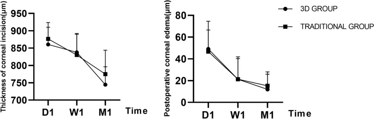
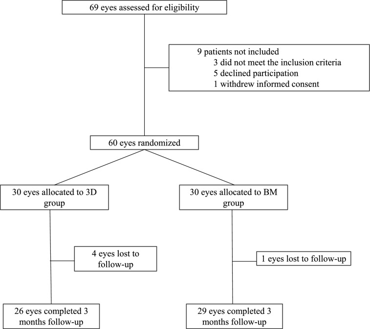
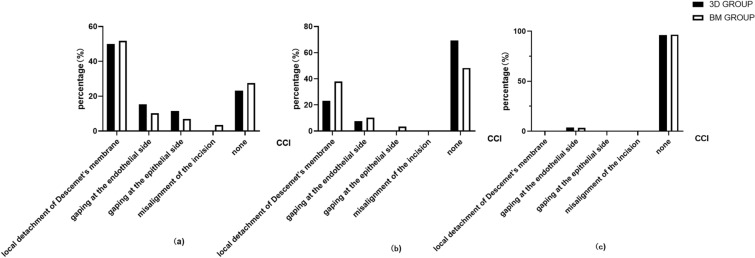
Of the 60 eyes randomly assigned between January 5, 2021, and May 9, 2021, 55 (26 eyes in the 3D group and 29 eyes in the BM group) were analyzed. The ECD loss rate was 8.1% in the 3D group and 12.3% in the BM group, but the difference was not statistically significant. Local detachment of Descemet's membrane was seen in 50% (13 eyes, 3D group) and 51.6% (15 eyes, BM group), wound gaping at the endothelial side in 15.4% (four eyes, 3D group) and 10.3% (four eyes, BM group), gaping at the epithelial side in 11.5% (three eyes, 3D group) and 6.9% (two eyes, BM group), and misalignment of the incision in 3.4% (one eye, BM group) 1 day after surgery. These abnormalities improved with time. There was no difference between the 3D group and BM group in terms of other ocular parameters or surgical parameters before and after surgery.

CONCLUSIONS

Using the 3D surgical system for phacoemulsification and IOL implantation surgery seems to result in similar ECD and CCI conditions as using a conventional binocular microscope.

TRIAL REGISTRATION

The protocol was registered on ClinicalTrials.gov (NCT04839250).

摘要

引言

比较三维(3D)可视化系统与传统双目显微镜(BM)在白内障超声乳化吸除联合人工晶状体植入手术中采用2.2毫米透明角膜切口(CCI)的手术效果。

方法

在这项随机对照临床研究中,60例年龄相关性白内障患者被分为两组,分别使用3D视觉系统(n = 30眼)(3D组)或双目显微镜(n = 30眼)(BM组)进行白内障手术。我们记录并统计分析了手术参数以及术前和术后的眼部参数。主要结局包括内皮细胞密度(ECD)的变化和CCI结构,次要结局包括其他眼部参数和手术参数。所有手术均遵循《赫尔辛基宣言》的原则。

结果

在2021年1月5日至2021年5月9日随机分配的60眼中,分析了55眼(3D组26眼,BM组29眼)。3D组的ECD损失率为8.1%,BM组为12.3%,但差异无统计学意义。3D组50%(13眼)和BM组51.6%(15眼)出现后弹力层局部脱离,3D组15.4%(4眼)和BM组10.3%(4眼)在内皮侧出现切口裂开,3D组11.5%(3眼)和BM组6.9%(2眼)在上皮侧出现切口裂开,术后1天BM组3.4%(1眼)出现切口错位。这些异常情况随时间改善。3D组和BM组在手术前后的其他眼部参数或手术参数方面无差异。

结论

在白内障超声乳化吸除联合人工晶状体植入手术中使用3D手术系统似乎与使用传统双目显微镜导致的ECD和CCI情况相似。

试验注册

该方案已在ClinicalTrials.gov(NCT04839250)上注册。

https://cdn.ncbi.nlm.nih.gov/pmc/blobs/b8d3/9253230/a188ff673866/40123_2022_537_Fig4_HTML.jpg
https://cdn.ncbi.nlm.nih.gov/pmc/blobs/b8d3/9253230/4f1e1c987515/40123_2022_537_Fig1_HTML.jpg
https://cdn.ncbi.nlm.nih.gov/pmc/blobs/b8d3/9253230/0a8e26f49694/40123_2022_537_Fig2_HTML.jpg
https://cdn.ncbi.nlm.nih.gov/pmc/blobs/b8d3/9253230/b1578f659696/40123_2022_537_Fig3_HTML.jpg
https://cdn.ncbi.nlm.nih.gov/pmc/blobs/b8d3/9253230/a188ff673866/40123_2022_537_Fig4_HTML.jpg

相似文献

1
Comparison of Three-Dimensional Surgical System Versus Binocular Microscope for Clear Corneal Incision in Cataract Surgery.白内障手术中三维手术系统与双目显微镜用于透明角膜切口的比较。
Ophthalmol Ther. 2022 Aug;11(4):1589-1600. doi: 10.1007/s40123-022-00537-4. Epub 2022 Jun 24.
2
Digital 3D "Heads-up" Cataract Surgery: Safety Profile and Comparison with the Conventional Microscope System.数字化 3D“抬头”白内障手术:安全性特征及与传统显微镜系统的比较。
Klin Monbl Augenheilkd. 2022 Aug;239(8):991-995. doi: 10.1055/a-1686-9124. Epub 2022 Feb 18.
3
Three-dimensional digital visualization of phacoemulsification and intraocular lens implantation.超声乳化白内障吸除术及人工晶状体植入的三维数字化可视化。
Indian J Ophthalmol. 2019 Mar;67(3):341-343. doi: 10.4103/ijo.IJO_1012_18.
4
Clear corneal incision architecture in the immediate postoperative period evaluated using optical coherence tomography.使用光学相干断层扫描评估术后即刻的透明角膜切口结构。
J Cataract Refract Surg. 2007 Aug;33(8):1429-35. doi: 10.1016/j.jcrs.2007.04.011.
5
Bimanual microincision cataract surgery with implantation of the new Incise MJ14 intraocular lens through a 1.4 mm incision.双手微切口白内障手术,通过1.4毫米切口植入新型Incise MJ14人工晶状体。
Int J Ophthalmol. 2017 Nov 18;10(11):1710-1715. doi: 10.18240/ijo.2017.11.12. eCollection 2017.
6
Intracameral air injection after completion of phacoemulsification cataract surgery: Evaluation of corneal incisions with optical coherence tomography.白内障超声乳化手术后前房内注入空气:用光学相干断层扫描评估角膜切口
J Curr Ophthalmol. 2019 Feb 13;31(2):142-149. doi: 10.1016/j.joco.2019.01.009. eCollection 2019 Jun.
7
Heads-up Cataract Surgery: Complication Rates, Surgical Duration, and Comparison With Traditional Microscopes.即时白内障手术:并发症发生率、手术持续时间及与传统显微镜的比较。
J Refract Surg. 2019 May 1;35(5):318-322. doi: 10.3928/1081597X-20190410-02.
8
Intraocular lens-shell technique: adjustment of the surgical procedure leads to greater safety when treating dense nuclear cataracts.人工晶状体囊袋技术:在治疗致密核性白内障时,调整手术操作可提高安全性。
PLoS One. 2014 Nov 17;9(11):e112663. doi: 10.1371/journal.pone.0112663. eCollection 2014.
9
Intraocular pressure and wound state immediately after long versus short clear corneal incision cataract surgery.长切口与短切口透明角膜切口白内障手术后即刻的眼压及伤口状态
Jpn J Ophthalmol. 2018 Nov;62(6):621-627. doi: 10.1007/s10384-018-0626-1. Epub 2018 Oct 6.
10
Refractive stability of a new single-piece hydrophobic acrylic intraocular lens and corneal wound repair after implantation using a new automated intraocular lens delivery system.新型疏水丙烯酸一体式人工晶状体的屈光度稳定性及应用新型自动人工晶状体推送系统行角膜伤口修复后的愈合情况。
PLoS One. 2020 Sep 2;15(9):e0238366. doi: 10.1371/journal.pone.0238366. eCollection 2020.

本文引用的文献

1
Early corneal pachymetry maps after cataract surgery and influence of 3D digital visualization system in minimizing corneal oedema.白内障手术后早期角膜生物力学地形图及其 3D 数字可视化系统对减轻角膜水肿的影响
Acta Ophthalmol. 2022 Aug;100(5):e1088-e1094. doi: 10.1111/aos.15060. Epub 2021 Nov 8.
2
Incidence of Incision-Related Descemet Membrane Detachment Using Phacoemulsification With Trapezoid vs Conventional 2.2-mm Clear Corneal Incision: A Randomized Clinical Trial.梯形与常规 2.2mm 透明角膜切口超声乳化术中切口相关的后弹力层脱离发生率:一项随机临床试验。
JAMA Ophthalmol. 2021 Nov 1;139(11):1228-1234. doi: 10.1001/jamaophthalmol.2021.4148.
3
Three-Dimensional Heads-up Cataract Surgery Using Femtosecond Laser: Efficiency, Efficacy, Safety, and Medical Education-A Randomized Clinical Trial.
飞秒激光辅助的三维眼前节白内障手术:效率、效果、安全性和医学教育——一项随机临床试验。
Transl Vis Sci Technol. 2021 Aug 2;10(9):4. doi: 10.1167/tvst.10.9.4.
4
Use of the heads-up NGENUITY 3D Visualization System for vitreoretinal surgery: a retrospective evaluation of outcomes in a French tertiary center.使用平视 NGENUITY 3D 可视化系统进行玻璃体视网膜手术:法国三级中心的回顾性结果评估。
Sci Rep. 2021 May 11;11(1):10031. doi: 10.1038/s41598-021-88993-z.
5
Heads-up 3D eye surgery: Safety outcomes and technological review after 2 years of day-to-day use.前瞻性3D眼科手术:日常使用两年后的安全性结果与技术综述
Eur J Ophthalmol. 2021 Apr 30:11206721211012856. doi: 10.1177/11206721211012856.
6
Contrast, visibility, and color balance between the microscope versus intracameral illumination in cataract surgery using a 3D visualization system.在使用 3D 可视化系统的白内障手术中,显微镜与眼内照明之间的对比度、可见度和色彩平衡。
Indian J Ophthalmol. 2021 Apr;69(4):927-931. doi: 10.4103/ijo.IJO_1825_20.
7
Three-dimensional heads up display in anterior segment surgeries- Expanding frontiers in the COVID-19 era.眼前节手术中的三维抬头显示器——新冠疫情时代的前沿拓展
Indian J Ophthalmol. 2020 Nov;68(11):2338-2340. doi: 10.4103/ijo.IJO_2978_20.
8
Heads-up Descemet Membrane Endothelial Keratoplasty Surgery: Feasibility, Surgical Duration, Complication Rates, and Comparison With a Conventional Microscope.顶压式 Descemet 膜内皮角膜移植手术:可行性、手术时间、并发症发生率,以及与传统显微镜的比较。
Cornea. 2021 Apr;40(4):415-419. doi: 10.1097/ICO.0000000000002419.
9
Comparison of microscopic illumination between a three-dimensional heads-up system and eyepiece in cataract surgery.三维头高位系统与目镜在白内障手术中显微镜照明的比较。
Eur J Ophthalmol. 2021 Jul;31(4):1817-1821. doi: 10.1177/1120672120929962. Epub 2020 Jun 8.
10
Comparing Heads-Up versus Binocular Microscope Visualization Systems in Anterior and Posterior Segment Surgeries: A Retrospective Study.头高位与双目显微镜可视化系统在前节和后节手术中的比较:一项回顾性研究。
Ophthalmologica. 2020;243(5):347-354. doi: 10.1159/000507088. Epub 2020 Mar 11.