Lindhardt Thomas Beck, Gutiérrez-Jiménez Eugenio, Liang Zhifeng, Hansen Brian
Department of Clinical Medicine, Center of Functionally Integrative Neuroscience, Aarhus University, Aarhus, Denmark.
CAS Center for Excellence in Brain Sciences and Intelligence Technology, Institute of Neuroscience, Chinese Academy of Sciences, Shanghai, China.
Front Neurosci. 2022 Jun 10;16:853527. doi: 10.3389/fnins.2022.853527. eCollection 2022.
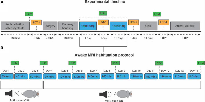
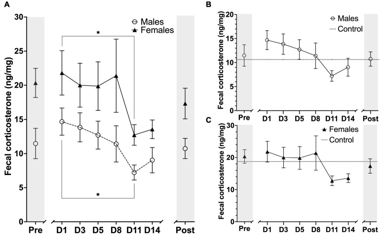
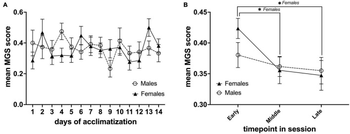
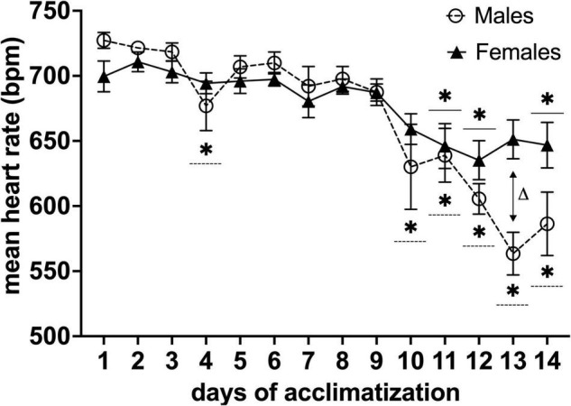
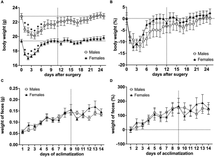
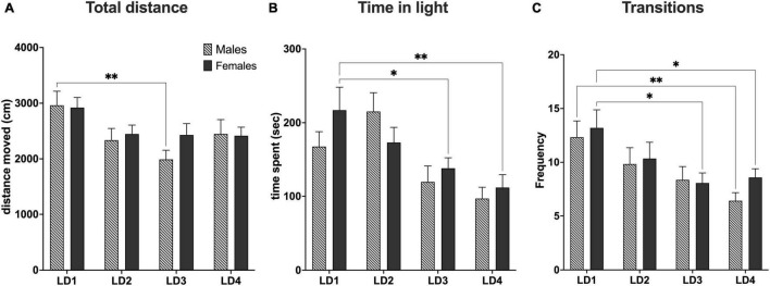
Traditionally, preclinical magnetic resonance imaging (MRI) has been performed in anesthetized animals. However, anesthesia has been shown to perturb normal brain function and physiology. Such effects limit our ability to detect subtle physiological alterations in disease models and treatment studies, thus hampering discovery and compromising generality of findings. Therefore, methods for awake animal MRI are needed to study the rodent brain in its natural physiological state, free of anesthetics. Current setups for awake animal MRI rely on restraining systems to avoid animal movement during scanning. To reduce restraint stress, animals are habituated to the scanner environment prior to MRI data collection. To date, however, most awake MRI studies employ male rodents only. This is a fundamental limitation as results obtained may be pertinent only to half of the population. We characterized training and habituation responses of male and female mice to provide improved, sex-dependent training procedures for awake mouse MRI. We recorded heart rate, monitored behavioral responses (body weight and weight) and fecal corticosterone levels (FCM) as indicators of wellbeing and stress during a 14-day progressive habituation protocol. In addition, we also assessed discomfort levels and anxiety using the mouse grimace scale (MGS) and light/dark test (LDT), respectively. All scores were compared between both groups. We found that heart rate was significantly decreased after 10 and 11 days of training for both males and females, respectively. However, the specific time course for this decrease was significantly different between males and females, and females exhibited higher anxiety levels during habituation and 14 days after habituation than males. Lastly, we also found that mean FCM levels for both groups were decreased after 11 days of MRI habituation. The present work shows that mice can be successfully trained for extended MRI sessions which is necessary for many (particularly non-fMRI) studies. Importantly, we find that males and females differ in their response to awake MRI habituation, which should be considered in future awake MRI studies that aim to include male and female mice.
传统上,临床前磁共振成像(MRI)是在麻醉的动物身上进行的。然而,已证明麻醉会干扰正常的脑功能和生理状态。这些影响限制了我们在疾病模型和治疗研究中检测细微生理变化的能力,从而阻碍了发现并损害了研究结果的普遍性。因此,需要清醒动物MRI方法来研究处于自然生理状态、未使用麻醉剂的啮齿动物大脑。当前的清醒动物MRI设置依赖于约束系统,以避免动物在扫描过程中移动。为了减轻约束压力,在收集MRI数据之前,让动物适应扫描仪环境。然而,迄今为止,大多数清醒MRI研究仅使用雄性啮齿动物。这是一个根本性的限制,因为所获得的结果可能仅适用于一半的群体。我们对雄性和雌性小鼠的训练和适应反应进行了表征,以提供改进的、性别依赖性的清醒小鼠MRI训练程序。在一个为期14天的渐进性适应方案中,我们记录心率、监测行为反应(体重和体重变化)以及粪便皮质酮水平(FCM),作为健康和压力的指标。此外,我们还分别使用小鼠 grimace量表(MGS)和明暗试验(LDT)评估不适水平和焦虑程度。比较了两组的所有分数。我们发现,雄性和雌性小鼠分别在训练10天和11天后心率显著下降。然而,这种下降的具体时间进程在雄性和雌性之间存在显著差异,并且雌性在适应过程中和适应后14天表现出比雄性更高的焦虑水平。最后,我们还发现,两组的平均FCM水平在MRI适应11天后均有所下降。目前的工作表明,小鼠可以成功地接受长时间MRI扫描训练,这对许多(特别是非功能磁共振成像)研究来说是必要的。重要的是,我们发现雄性和雌性对清醒MRI适应的反应不同,这在未来旨在纳入雄性和雌性小鼠的清醒MRI研究中应予以考虑。