• 文献检索
  • 文档翻译
  • 深度研究
  • 学术资讯
  • Suppr Zotero 插件Zotero 插件
  • 邀请有礼
  • 套餐&价格
  • 历史记录
应用&插件
Suppr Zotero 插件Zotero 插件浏览器插件Mac 客户端Windows 客户端微信小程序
定价
高级版会员购买积分包购买API积分包
服务
文献检索文档翻译深度研究API 文档MCP 服务
关于我们
关于 Suppr公司介绍联系我们用户协议隐私条款
关注我们

Suppr 超能文献

核心技术专利:CN118964589B侵权必究
粤ICP备2023148730 号-1Suppr @ 2026

文献检索

告别复杂PubMed语法,用中文像聊天一样搜索,搜遍4000万医学文献。AI智能推荐,让科研检索更轻松。

立即免费搜索

文件翻译

保留排版,准确专业,支持PDF/Word/PPT等文件格式,支持 12+语言互译。

免费翻译文档

深度研究

AI帮你快速写综述,25分钟生成高质量综述,智能提取关键信息,辅助科研写作。

立即免费体验

术前骨盆 MRI 和 2-[F]FDG PET/CT 用于子宫内膜癌的淋巴结分期和预后预测——是否需要重新审视当前的影像学指南?

Preoperative pelvic MRI and 2-[F]FDG PET/CT for lymph node staging and prognostication in endometrial cancer-time to revisit current imaging guidelines?

机构信息

Mohn Medical Imaging and Visualization Centre (MMIV), Department of Radiology, Haukeland University Hospital, Bergen, Norway.

Section for Radiology, Department of Clinical Medicine, University of Bergen, Bergen, Norway.

出版信息

Eur Radiol. 2023 Jan;33(1):221-232. doi: 10.1007/s00330-022-08949-3. Epub 2022 Jun 28.

DOI:10.1007/s00330-022-08949-3
PMID:35763096
原文链接:https://pmc.ncbi.nlm.nih.gov/articles/PMC9755079/
Abstract

OBJECTIVE

This study presents the diagnostic performance of four different preoperative imaging workups (IWs) for prediction of lymph node metastases (LNMs) in endometrial cancer (EC): pelvic MRI alone (IW1), MRI and [F]FDG-PET/CT in all patients (IW2), MRI with selective [F]FDG-PET/CT if high-risk preoperative histology (IW3), and MRI with selective [F]FDG-PET/CT if MRI indicates FIGO stage ≥ 1B (IW4).

METHODS

In 361 EC patients, preoperative staging parameters from both pelvic MRI and [F]FDG-PET/CT were recorded. Area under receiver operating characteristic curves (ROC AUC) compared the diagnostic performance for the different imaging parameters and workups for predicting surgicopathological FIGO stage. Survival data were assessed using Kaplan-Meier estimator with log-rank test.

RESULTS

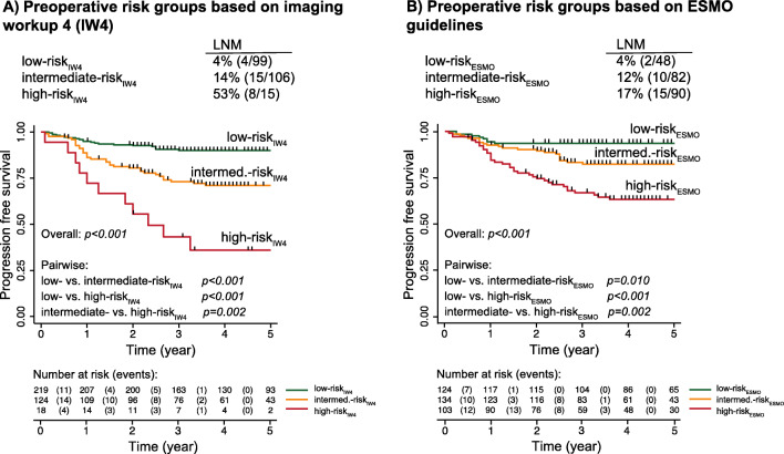
MRI and [F]FDG-PET/CT staging parameters yielded similar AUCs for predicting corresponding FIGO staging parameters in low-risk versus high-risk histology groups (p ≥ 0.16). The sensitivities, specificities, and AUCs for LNM prediction were as follows: IW1-33% [9/27], 95% [185/193], and 0.64; IW2-56% [15/27], 90% [174/193], and 0.73 (p = 0.04 vs. IW1); IW3-44% [12/27], 94% [181/193], and 0.69 (p = 0.13 vs. IW1); and IW4-52% [14/27], 91% [176/193], and 0.72 (p = 0.06 vs. IW1). IW3 and IW4 selected 34% [121/361] and 54% [194/361] to [F]FDG-PET/CT, respectively. Employing IW4 identified three distinct patient risk groups that exhibited increasing FIGO stage (p < 0.001) and stepwise reductions in survival (p ≤ 0.002).

CONCLUSION

Selective [F]FDG-PET/CT in patients with high-risk MRI findings yields better detection of LNM than MRI alone, and similar diagnostic performance to that of MRI and [F]FDG-PET/CT in all.

KEY POINTS

• Imaging by MRI and [F]FDG PET/CT yields similar diagnostic performance in low- and high-risk histology groups for predicting central FIGO staging parameters. • Utilizing a stepwise imaging workup with MRI in all patients and [F]FDG-PET/CT in selected patients based on MRI findings identifies preoperative risk groups exhibiting significantly different survival. • The proposed imaging workup selecting ~54% of the patients to [F]FDG-PET/CT yield better detection of LNMs than MRI alone, and similar LNM detection to that of MRI and [F]FDG-PET/CT in all.

摘要

目的

本研究提出了四种不同术前影像学检查(IW)在预测子宫内膜癌(EC)淋巴结转移(LNM)中的诊断性能:盆腔 MRI 单独(IW1)、所有患者的 MRI 和 [F]FDG-PET/CT(IW2)、如果术前组织学为高危,则进行 MRI 和选择性 [F]FDG-PET/CT(IW3)、如果 MRI 提示 FIGO 分期≥1B,则进行 MRI 和选择性 [F]FDG-PET/CT(IW4)。

方法

在 361 例 EC 患者中,记录了盆腔 MRI 和 [F]FDG-PET/CT 的术前分期参数。受试者工作特征曲线(ROC AUC)下的面积比较了不同成像参数和 IW 对预测手术病理 FIGO 分期的诊断性能。使用 Kaplan-Meier 估计器和对数秩检验评估生存数据。

结果

MRI 和 [F]FDG-PET/CT 分期参数在低风险与高风险组织学组中预测相应的 FIGO 分期参数时具有相似的 AUC(p≥0.16)。LNM 预测的灵敏度、特异性和 AUC 如下:IW1-33%[9/27]、95%[185/193]和 0.64;IW2-56%[15/27]、90%[174/193]和 0.73(p=0.04 比 IW1);IW3-44%[12/27]、94%[181/193]和 0.69(p=0.13 比 IW1);IW4-52%[14/27]、91%[176/193]和 0.72(p=0.06 比 IW1)。IW3 和 IW4 分别将 34%[121/361]和 54%[194/361]的患者选择至 [F]FDG-PET/CT。采用 IW4 可识别出三个不同的患者风险组,这些组表现出 FIGO 分期的递增(p<0.001)和生存率的逐步降低(p≤0.002)。

结论

在高危 MRI 发现的患者中进行选择性 [F]FDG-PET/CT 比单独 MRI 更能检测到 LNM,且与所有患者的 MRI 和 [F]FDG-PET/CT 具有相似的诊断性能。

关键点

  1. MRI 和 [F]FDG-PET/CT 成像在预测中央 FIGO 分期参数方面,在低风险和高风险组织学组中具有相似的诊断性能。

  2. 采用逐步成像工作流程,所有患者均进行 MRI,根据 MRI 结果选择高危患者进行 [F]FDG-PET/CT,可识别出具有显著不同生存率的术前风险组。

  3. 选择约 54%的患者进行 [F]FDG-PET/CT 的建议成像工作流程,比单独 MRI 更能检测到 LNM,且与所有患者的 MRI 和 [F]FDG-PET/CT 具有相似的 LNM 检测性能。

https://cdn.ncbi.nlm.nih.gov/pmc/blobs/3c7e/9755079/e8c3f928af39/330_2022_8949_Fig3_HTML.jpg
https://cdn.ncbi.nlm.nih.gov/pmc/blobs/3c7e/9755079/bc03e9f1077f/330_2022_8949_Fig1_HTML.jpg
https://cdn.ncbi.nlm.nih.gov/pmc/blobs/3c7e/9755079/8f4e2ed2c311/330_2022_8949_Fig2_HTML.jpg
https://cdn.ncbi.nlm.nih.gov/pmc/blobs/3c7e/9755079/e8c3f928af39/330_2022_8949_Fig3_HTML.jpg
https://cdn.ncbi.nlm.nih.gov/pmc/blobs/3c7e/9755079/bc03e9f1077f/330_2022_8949_Fig1_HTML.jpg
https://cdn.ncbi.nlm.nih.gov/pmc/blobs/3c7e/9755079/8f4e2ed2c311/330_2022_8949_Fig2_HTML.jpg
https://cdn.ncbi.nlm.nih.gov/pmc/blobs/3c7e/9755079/e8c3f928af39/330_2022_8949_Fig3_HTML.jpg

相似文献

1
Preoperative pelvic MRI and 2-[F]FDG PET/CT for lymph node staging and prognostication in endometrial cancer-time to revisit current imaging guidelines?术前骨盆 MRI 和 2-[F]FDG PET/CT 用于子宫内膜癌的淋巴结分期和预后预测——是否需要重新审视当前的影像学指南?
Eur Radiol. 2023 Jan;33(1):221-232. doi: 10.1007/s00330-022-08949-3. Epub 2022 Jun 28.
2
Preoperative 18F-FDG PET/CT tumor markers outperform MRI-based markers for the prediction of lymph node metastases in primary endometrial cancer.术前 18F-FDG PET/CT 肿瘤标志物预测原发性子宫内膜癌淋巴结转移的效能优于 MRI 标志物。
Eur Radiol. 2020 May;30(5):2443-2453. doi: 10.1007/s00330-019-06622-w. Epub 2020 Feb 7.
3
Lymph Node Staging with a Combined Protocol of F-FDG PET/MRI and Sentinel Node SPECT/CT: A Prospective Study in Patients with FIGO I/II Cervical Carcinoma.基于 F-FDG PET/MRI 联合前哨淋巴结 SPECT/CT 方案的淋巴结分期:FIGO I/II 期宫颈癌患者的前瞻性研究。
J Nucl Med. 2021 Aug 1;62(8):1062-1067. doi: 10.2967/jnumed.120.255919. Epub 2021 Jan 28.
4
Preoperative lymph-node staging of invasive urothelial bladder cancer with 18F-fluorodeoxyglucose positron emission tomography/computed axial tomography and magnetic resonance imaging: correlation with histopathology.18F-氟脱氧葡萄糖正电子发射断层扫描/计算机断层扫描和磁共振成像对浸润性膀胱尿路上皮癌的术前淋巴结分期:与组织病理学的相关性
Scand J Urol Nephrol. 2011 Mar;45(2):122-8. doi: 10.3109/00365599.2010.544672. Epub 2011 Jan 13.
5
The Value of Preoperative Positron Emission Tomography/Computed Tomography in Node-Negative Endometrial Cancer on Magnetic Resonance Imaging.术前正电子发射断层扫描/计算机断层扫描在磁共振成像阴性淋巴结子宫内膜癌中的价值。
Ann Surg Oncol. 2017 Aug;24(8):2303-2310. doi: 10.1245/s10434-017-5901-8. Epub 2017 May 26.
6
Diagnostic Value of F-fluorodeoxyglucose Positron Emission Tomography with Computed Tomography for Lymph Node Staging in Patients with Upper Tract Urothelial Carcinoma.F-氟代脱氧葡萄糖正电子发射断层扫描与计算机断层扫描对肾盂尿路上皮癌患者淋巴结分期的诊断价值。
Eur Urol Oncol. 2020 Feb;3(1):73-79. doi: 10.1016/j.euo.2019.09.004. Epub 2019 Oct 5.
7
Diagnostic value of integrated F-FDG PET/MRI for staging of endometrial carcinoma: comparison with PET/CT.F-FDG PET/MRI 对子宫内膜癌分期的诊断价值:与 PET/CT 的比较。
BMC Cancer. 2022 Sep 1;22(1):947. doi: 10.1186/s12885-022-10037-0.
8
Comparison of FDG PET/CT and MRI in lymph node staging of endometrial cancer.18F-氟脱氧葡萄糖正电子发射断层扫描/计算机断层扫描(FDG PET/CT)与磁共振成像(MRI)在子宫内膜癌淋巴结分期中的比较
Ann Nucl Med. 2016 Feb;30(2):104-13. doi: 10.1007/s12149-015-1037-8. Epub 2015 Nov 6.
9
The Role of Preoperative Imaging in the Detection of Lateral Lymph Node Metastases in Rectal Cancer: A Systematic Review and Diagnostic Test Meta-analysis.术前影像学在直肠癌侧方淋巴结转移检测中的作用:系统评价和诊断试验荟萃分析。
Dis Colon Rectum. 2022 Dec 1;65(12):1436-1446. doi: 10.1097/DCR.0000000000002537. Epub 2022 Aug 29.
10
Prognostic value of metabolic tumor volume and total lesion glycolysis from ¹⁸F-FDG PET/CT in lymph node metastases and risk stratification of endometrial carcinoma.¹⁸F-FDG PET/CT 代谢肿瘤体积和总肿瘤糖酵解与子宫内膜癌淋巴结转移及危险分层的预后价值。
J Gynecol Oncol. 2019 Nov;30(6):e89. doi: 10.3802/jgo.2019.30.e89.

引用本文的文献

1
Molecular Imaging in Endometrial Cancer: A Narrative Review.子宫内膜癌的分子成像:一篇叙述性综述
Cancers (Basel). 2025 Aug 8;17(16):2608. doi: 10.3390/cancers17162608.
2
Diagnostic Accuracy and Interrater Agreement of FDG-PET/CT Lymph Node Staging in High-Risk Endometrial Cancer: The SENTIREC-Endo Study.18F-氟脱氧葡萄糖正电子发射断层扫描/计算机断层扫描(FDG-PET/CT)对高危子宫内膜癌淋巴结分期的诊断准确性及评分者间一致性:SENTIREC-Endo研究
Cancers (Basel). 2025 Jul 19;17(14):2396. doi: 10.3390/cancers17142396.
3
Contrast-enhanced MRI and CT in evaluating treatment response for recurrent endometrial cancer: a retrospective case-control study.

本文引用的文献

1
Global Cancer Statistics 2020: GLOBOCAN Estimates of Incidence and Mortality Worldwide for 36 Cancers in 185 Countries.《全球癌症统计数据 2020:全球 185 个国家和地区 36 种癌症的发病率和死亡率估计》。
CA Cancer J Clin. 2021 May;71(3):209-249. doi: 10.3322/caac.21660. Epub 2021 Feb 4.
2
ESGO/ESTRO/ESP guidelines for the management of patients with endometrial carcinoma.ESGO/ESTRO/ESP 子宫内膜癌管理指南。
Int J Gynecol Cancer. 2021 Jan;31(1):12-39. doi: 10.1136/ijgc-2020-002230. Epub 2020 Dec 18.
3
Maintained survival outcome after reducing lymphadenectomy rates and optimizing adjuvant treatment in endometrial cancer.
对比增强磁共振成像和计算机断层扫描在评估复发性子宫内膜癌治疗反应中的应用:一项回顾性病例对照研究
Am J Cancer Res. 2025 May 15;15(5):2077-2096. doi: 10.62347/TUED9082. eCollection 2025.
4
Is presumed clinical stage I endometrial cancer using PET-CT and MRI accurate in predicting surgical staging?使用正电子发射断层扫描-计算机断层扫描(PET-CT)和磁共振成像(MRI)推断的临床I期子宫内膜癌在预测手术分期方面准确吗?
J Gynecol Oncol. 2025 Mar;36(2):e25. doi: 10.3802/jgo.2025.36.e25. Epub 2025 Jan 7.
5
Both intra- and peri-tumoral radiomics signatures can be used to predict lymphatic vascular space invasion and lymphatic metastasis positive status from endometrial cancer MR imaging.肿瘤内和肿瘤周围放射组学特征均可用于预测子宫内膜癌 MRI 中的淋巴管侵犯和淋巴管转移阳性状态。
Abdom Radiol (NY). 2024 Nov;49(11):4140-4150. doi: 10.1007/s00261-024-04432-3. Epub 2024 Jun 25.
6
Endometrial Cancer: 2023 Revised FIGO Staging System and the Role of Imaging.子宫内膜癌:2023年修订的国际妇产科联盟(FIGO)分期系统及影像学的作用
Cancers (Basel). 2024 May 14;16(10):1869. doi: 10.3390/cancers16101869.
7
Staging by imaging in gynecologic cancer and the role of ultrasound: an update of European joint consensus statements.妇科癌症的影像学分期和超声的作用:欧洲联合共识声明的更新。
Int J Gynecol Cancer. 2024 Mar 4;34(3):363-378. doi: 10.1136/ijgc-2023-004609.
8
Preoperative CA125 Significantly Improves Risk Stratification in High-Grade Endometrial Cancer.术前CA125显著改善高级别子宫内膜癌的风险分层。
Cancers (Basel). 2023 May 4;15(9):2605. doi: 10.3390/cancers15092605.
在降低淋巴结切除术率和优化子宫内膜癌辅助治疗后,保持生存结果。
Gynecol Oncol. 2021 Feb;160(2):396-404. doi: 10.1016/j.ygyno.2020.12.002. Epub 2020 Dec 13.
4
Endometrial Cancer.子宫内膜癌
N Engl J Med. 2020 Nov 19;383(21):2053-2064. doi: 10.1056/NEJMra1514010.
5
Whole-Volume Tumor MRI Radiomics for Prognostic Modeling in Endometrial Cancer.全容积肿瘤 MRI 影像组学在子宫内膜癌预后建模中的应用。
J Magn Reson Imaging. 2021 Mar;53(3):928-937. doi: 10.1002/jmri.27444. Epub 2020 Nov 16.
6
Preoperative risk stratification in endometrial cancer (ENDORISK) by a Bayesian network model: A development and validation study.基于贝叶斯网络模型的子宫内膜癌术前风险分层(ENDORISK):一项开发和验证研究。
PLoS Med. 2020 May 15;17(5):e1003111. doi: 10.1371/journal.pmed.1003111. eCollection 2020 May.
7
Development of prediction models for lymph node metastasis in endometrioid endometrial carcinoma.预测子宫内膜样腺癌淋巴结转移的模型的建立。
Br J Cancer. 2020 Mar;122(7):1014-1022. doi: 10.1038/s41416-020-0745-6. Epub 2020 Feb 10.
8
Preoperative 18F-FDG PET/CT tumor markers outperform MRI-based markers for the prediction of lymph node metastases in primary endometrial cancer.术前 18F-FDG PET/CT 肿瘤标志物预测原发性子宫内膜癌淋巴结转移的效能优于 MRI 标志物。
Eur Radiol. 2020 May;30(5):2443-2453. doi: 10.1007/s00330-019-06622-w. Epub 2020 Feb 7.
9
F-FDG PET/CT Quantitative Parameters and Texture Analysis Effectively Differentiate Endometrial Precancerous Lesion and Early-Stage Carcinoma.F-FDG PET/CT定量参数及纹理分析可有效鉴别子宫内膜癌前病变与早期癌。
Mol Imaging. 2019 Jan-Dec;18:1536012119856965. doi: 10.1177/1536012119856965.
10
Current recommendations and recent progress in endometrial cancer.子宫内膜癌的当前建议和最新进展。
CA Cancer J Clin. 2019 Jul;69(4):258-279. doi: 10.3322/caac.21561. Epub 2019 May 10.