Leibniz-Forschungsinstitut für Molekulare Pharmakologie (FMP) and Max-Delbrück-Centrum für Molekulare Medizin (MDC), Berlin, Germany.
Integrative Metabolomics and Proteomics, Berlin Institute of Medical Systems Biology/Max-Delbrück-Centrum für Molekulare Medizin, Berlin, Germany.
J Am Soc Nephrol. 2022 Aug;33(8):1528-1545. doi: 10.1681/ASN.2021111458. Epub 2022 Jul 1.
Volume-regulated anion channels (VRACs) are heterohexamers of LRRC8A with LRRC8B, -C, -D, or -E in various combinations. Depending on the subunit composition, these swelling-activated channels conduct chloride, amino acids, organic osmolytes, and drugs. Despite VRACs' role in cell volume regulation, and large osmolarity changes in the kidney, neither the localization nor the function of VRACs in the kidney is known.
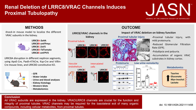
Mice expressing epitope-tagged LRRC8 subunits were used to determine the renal localization of all VRAC subunits. Mice carrying constitutive deletions of -, or with inducible or cell-specific ablation of , were analyzed to assess renal functions of VRACs. Analysis included histology, urine and serum parameters in different diuresis states, and metabolomics.
The kidney expresses all five VRAC subunits with strikingly distinct localization. Whereas LRRC8C is exclusively found in vascular endothelium, all other subunits are found in the nephron. LRRC8E is specific for intercalated cells, whereas LRRC8A, LRRC8B, and LRRC8D are prominent in basolateral membranes of proximal tubules. Conditional deletion of LRRC8A in proximal but not distal tubules and constitutive deletion of LRRC8D cause proximal tubular injury, increased diuresis, and mild Fanconi-like symptoms.
VRAC/LRRC8 channels are crucial for the function and integrity of proximal tubules, but not for more distal nephron segments despite their larger need for volume regulation. LRRC8A/D channels may be required for the basolateral exit of many organic compounds, including cellular metabolites, in proximal tubules. Proximal tubular injury likely results from combined accumulation of several transported molecules in the absence of VRAC channels.
体积调节阴离子通道(VRAC)是由 LRRC8A 与 LRRC8B、-C、-D 或 -E 以各种组合形成的异六聚体。根据亚基组成的不同,这些肿胀激活的通道可传导氯离子、氨基酸、有机渗透物和药物。尽管 VRAC 在细胞体积调节和肾脏中的渗透压变化中起作用,但 VRAC 在肾脏中的定位和功能尚不清楚。
使用表达表位标记的 LRRC8 亚基的小鼠来确定所有 VRAC 亚基在肾脏中的定位。分析携带 - 组成型缺失或诱导型或细胞特异性消融的小鼠,以评估 VRAC 在肾脏中的功能。分析包括组织学、不同利尿状态下的尿液和血清参数以及代谢组学。
肾脏表达所有五个具有明显不同定位的 VRAC 亚基。虽然 LRRC8C 仅存在于血管内皮细胞中,但所有其他亚基都存在于肾单位中。LRRC8E 是间充质细胞特异性的,而 LRRC8A、LRRC8B 和 LRRC8D 则在近端肾小管的基底外侧膜中显著表达。LRRC8A 在近端肾小管而非远端肾小管中的条件性缺失以及 LRRC8D 的组成型缺失导致近端肾小管损伤、利尿增加和轻度 Fanconi 样症状。
VRAC/LRRC8 通道对于近端肾小管的功能和完整性至关重要,但对于更远端的肾单位并非如此,尽管它们对体积调节的需求更大。LRRC8A/D 通道可能是许多有机化合物(包括细胞代谢物)在近端肾小管中基底外侧出口所必需的。在没有 VRAC 通道的情况下,几种转运分子的累积可能导致近端肾小管损伤。