• 文献检索
  • 文档翻译
  • 深度研究
  • 学术资讯
  • Suppr Zotero 插件Zotero 插件
  • 邀请有礼
  • 套餐&价格
  • 历史记录
应用&插件
Suppr Zotero 插件Zotero 插件浏览器插件Mac 客户端Windows 客户端微信小程序
定价
高级版会员购买积分包购买API积分包
服务
文献检索文档翻译深度研究API 文档MCP 服务
关于我们
关于 Suppr公司介绍联系我们用户协议隐私条款
关注我们

Suppr 超能文献

核心技术专利:CN118964589B侵权必究
粤ICP备2023148730 号-1Suppr @ 2026

文献检索

告别复杂PubMed语法,用中文像聊天一样搜索,搜遍4000万医学文献。AI智能推荐,让科研检索更轻松。

立即免费搜索

文件翻译

保留排版,准确专业,支持PDF/Word/PPT等文件格式,支持 12+语言互译。

免费翻译文档

深度研究

AI帮你快速写综述,25分钟生成高质量综述,智能提取关键信息,辅助科研写作。

立即免费体验

上颌部分缺牙患者中数字化和传统印模的体外精度。

In vitro accuracy of digital and conventional impressions in the partially edentulous maxilla.

机构信息

Department of Prosthetic Dentistry, Heidelberg University Hospital, University of Heidelberg, Im Neuenheimer Feld 400, 69120, Heidelberg, Germany.

Institute of Medical Biometry, Heidelberg University Hospital, University of Heidelberg, Heidelberg, Germany.

出版信息

Clin Oral Investig. 2022 Nov;26(11):6491-6502. doi: 10.1007/s00784-022-04598-4. Epub 2022 Jul 1.

DOI:10.1007/s00784-022-04598-4
PMID:35778534
原文链接:https://pmc.ncbi.nlm.nih.gov/articles/PMC9643200/
Abstract

OBJECTIVES

This in vitro study compared the dimensional accuracy of conventional impressions (CI) with that of digital impressions (DI) in a partially edentulous maxilla. DIs were made by two intraoral scanners, Omnicam (OC) and Primescan (PS).

MATERIALS AND METHODS

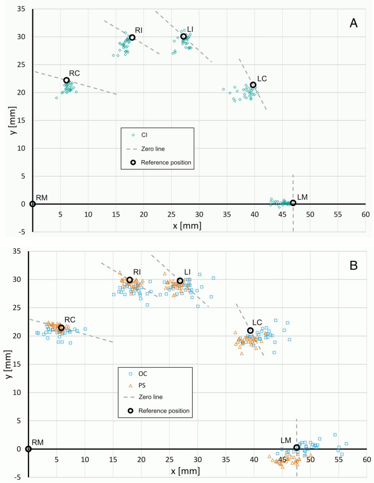
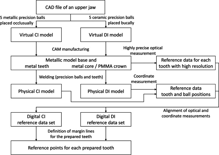
CI and both intraoral scanners were used to take 30 impressions of two identical reference models. CIs were poured with type 4 gypsum and the saw-cut models were digitized. The reference models simulated a maxilla with six prepared teeth that accommodated a cross-arch fixed partial denture. Center points of five precision balls and center points at the margin level of each prepared tooth were used to detect changes in dimensions and tooth axis between the reference model and the scans.

RESULTS

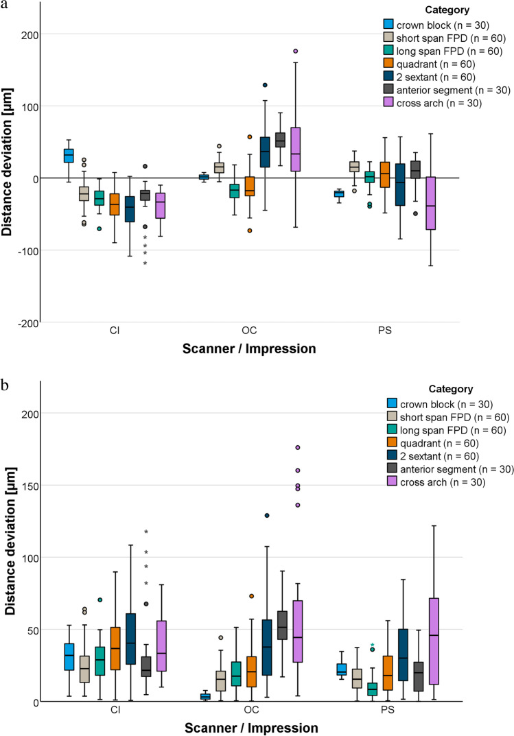
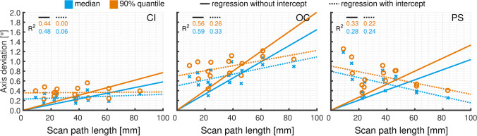
For DI, the largest deviations (176 µm for OC and 122 µm for PS) occurred over the cross-arch. For CI, the largest deviation (118 µm) occurred over the anterior segment. For shorter distances up to a quadrant, DI was superior to CI. For longer scan distances, DI was comparable (2 sextant and anterior segment) or inferior (cross-arch) to CI. Vertical and tooth axis deviations were significantly smaller for CI than for DI (p < 0.001).

CONCLUSIONS

The impression method affected the impression accuracy of a partially edentulous maxilla with prepared teeth. DI is recommended for scans up to a quadrant. Larger scan volumes are not yet suitable for fabricating a fixed partial denture because of the high scatter of accuracy values.

CLINICAL RELEVANCE

In contrast to conventional impressions, digital impressions lead to comparable or better results concerning scans up to a quadrant. Consequently, for larger scan volumes, several smaller scans should be performed or, if restoration-related not possible, it is recommended to take conventional impressions.

摘要

目的

本体外研究比较了上颌部分缺牙患者常规印模(CI)和数字印模(DI)的尺寸精度。DI 由两种口内扫描仪 Omnicam(OC)和 Primescan(PS)制作。

材料和方法

使用 CI 和两种口内扫描仪对两个相同的参考模型进行 30 次印模。CI 用 4 型石膏浇铸,对锯切模型进行数字化。参考模型模拟了一个上颌,有六颗预备牙,容纳了一个跨弓固定局部义齿。使用五个精密球的中心点和每个预备牙的边缘水平的中心点来检测参考模型和扫描之间的尺寸和牙轴变化。

结果

对于 DI,最大偏差(OC 为 176μm,PS 为 122μm)发生在跨弓上。对于 CI,最大偏差(118μm)发生在前部。对于短距离(至一个象限),DI 优于 CI。对于较长的扫描距离,DI 与 CI 相当(2 个六分仪和前部)或不如 CI(跨弓)。CI 的垂直和牙轴偏差明显小于 DI(p<0.001)。

结论

印模方法对上颌有预备牙的部分缺牙患者的印模精度有影响。建议在扫描至一个象限时使用 DI。由于准确性值的较大离散度,更大的扫描体积目前还不适合制作固定局部义齿。

临床意义

与传统印模相比,数字印模在扫描至一个象限时可以获得类似或更好的结果。因此,对于更大的扫描体积,应该进行几次较小的扫描,或者如果修复相关不可行,则建议进行传统印模。

https://cdn.ncbi.nlm.nih.gov/pmc/blobs/2e36/9643200/15c5a1f503d7/784_2022_4598_Fig6_HTML.jpg
https://cdn.ncbi.nlm.nih.gov/pmc/blobs/2e36/9643200/0fb0e95efc01/784_2022_4598_Fig1_HTML.jpg
https://cdn.ncbi.nlm.nih.gov/pmc/blobs/2e36/9643200/499d7f70ce14/784_2022_4598_Fig2_HTML.jpg
https://cdn.ncbi.nlm.nih.gov/pmc/blobs/2e36/9643200/b47db6fd393f/784_2022_4598_Fig3_HTML.jpg
https://cdn.ncbi.nlm.nih.gov/pmc/blobs/2e36/9643200/8aa95c1976a2/784_2022_4598_Fig4_HTML.jpg
https://cdn.ncbi.nlm.nih.gov/pmc/blobs/2e36/9643200/e66737afb8f5/784_2022_4598_Fig5_HTML.jpg
https://cdn.ncbi.nlm.nih.gov/pmc/blobs/2e36/9643200/15c5a1f503d7/784_2022_4598_Fig6_HTML.jpg

相似文献

1
In vitro accuracy of digital and conventional impressions in the partially edentulous maxilla.上颌部分缺牙患者中数字化和传统印模的体外精度。
Clin Oral Investig. 2022 Nov;26(11):6491-6502. doi: 10.1007/s00784-022-04598-4. Epub 2022 Jul 1.
2
Accuracy of complete- and partial-arch impressions of actual intraoral scanning systems in vitro.体外实际口腔内扫描系统全牙弓和部分牙弓印模的准确性。
Int J Comput Dent. 2019;22(1):11-19.
3
In-vitro accuracy of complete arch scans of the fully dentate and the partially edentulous maxilla.全牙列和部分牙列上颌的全弓扫描的体外准确性。
J Prosthodont Res. 2022 Oct 7;66(4):538-545. doi: 10.2186/jpr.JPR_D_21_00100. Epub 2021 Dec 7.
4
Effect of scan-path length on the scanning accuracy of completely dentate and partially edentulous maxillae.扫描路径长度对完全无牙颌和部分无牙颌上颌骨扫描精度的影响。
J Prosthet Dent. 2024 Jan;131(1):146-154. doi: 10.1016/j.prosdent.2022.02.016. Epub 2022 Mar 30.
5
Accuracy of impression-making methods in edentulous arches: An in vitro study encompassing conventional and digital methods.无牙颌印模方法的准确性:一项包含传统和数字方法的体外研究。
J Prosthet Dent. 2022 Sep;128(3):479-486. doi: 10.1016/j.prosdent.2020.09.038. Epub 2021 Feb 11.
6
Accuracy of full-arch digital impressions: an in vitro and in vivo comparison.全口数字化印模的准确性:一项体外与体内比较研究。
Clin Oral Investig. 2020 Feb;24(2):735-745. doi: 10.1007/s00784-019-02965-2. Epub 2019 May 27.
7
Accuracy of Intraoral Digital Impressions for Whole Upper Jaws, Including Full Dentitions and Palatal Soft Tissues.全上颌(包括完整牙列和腭部软组织)口内数字化印模的准确性
PLoS One. 2016 Jul 6;11(7):e0158800. doi: 10.1371/journal.pone.0158800. eCollection 2016.
8
Accuracy of complete-arch dental impressions: a new method of measuring trueness and precision.全牙弓口腔印模的准确性:一种测量准确性和精密度的新方法。
J Prosthet Dent. 2013 Feb;109(2):121-8. doi: 10.1016/S0022-3913(13)60028-1.
9
A new method for assessing the accuracy of full arch impressions in patients.一种评估全牙弓印模准确性的新方法。
J Dent. 2016 Dec;55:68-74. doi: 10.1016/j.jdent.2016.10.002. Epub 2016 Oct 4.
10
Accuracy of digital complete-arch, multi-implant scans made in the edentulous jaw with gingival movement simulation: An in vitro study.无牙颌中使用牙龈运动模拟进行的数字化全颌、多种植体扫描的准确性:一项体外研究。
J Prosthet Dent. 2022 Sep;128(3):468-478. doi: 10.1016/j.prosdent.2020.12.037. Epub 2021 Feb 19.

引用本文的文献

1
In-vitro validation of a new method to assess the clinical accuracy of complete arch impressions.一种评估全牙弓印模临床准确性的新方法的体外验证
Clin Oral Investig. 2025 Feb 25;29(3):156. doi: 10.1007/s00784-025-06236-1.
2
Dimensional Accuracy of Novel Vinyl Polysiloxane Compared with Polyether Impression Materials: An In Vitro Study.新型乙烯基聚硅氧烷与聚醚印模材料的尺寸精度:一项体外研究
Materials (Basel). 2024 Aug 27;17(17):4221. doi: 10.3390/ma17174221.
3
Dimensional Accuracy of Different Three-Dimensional Printing Models as a Function of Varying the Printing Parameters.

本文引用的文献

1
Use of measuring gauges for accuracy analysis of intraoral scanners: a pilot study.使用测量规进行口腔内扫描仪的准确性分析:一项初步研究。
J Adv Prosthodont. 2021 Aug;13(4):191-204. doi: 10.4047/jap.2021.13.4.191. Epub 2021 Aug 26.
2
Evaluation of trueness and precision of two intraoral scanners and a conventional impression: an in vivo clinical study.两种口内扫描仪和传统印模的准确性和精密度评估:一项体内临床研究。
Quintessence Int. 2021 Oct 19;52(10):904-910. doi: 10.3290/j.qi.b1901329.
3
Trueness and precision of intraoral scanners in the maxillary dental arch: an in vivo analysis.
不同三维打印模型的尺寸精度随打印参数变化的函数关系
Materials (Basel). 2024 Jul 22;17(14):3616. doi: 10.3390/ma17143616.
4
Comparative Accuracy of Intraoral and Extraoral Digital Workflows for Short Span Implant Supported Fixed Partial Denture Fabrication: An In Vitro Study.口内和口外数字化工作流程在短跨度种植体支持的固定局部义齿制作中的准确性比较:一项体外研究。
Med Sci Monit. 2024 Mar 19;30:e943706. doi: 10.12659/MSM.943706.
5
Accuracy of Computerized Optical Impression Making in Fabrication of Removable Dentures for Partially Edentulous Jaws: An In Vivo Feasibility Study.计算机光学印模制作在部分无牙颌可摘局部义齿修复中的准确性:一项体内可行性研究。
J Funct Biomater. 2023 Sep 5;14(9):458. doi: 10.3390/jfb14090458.
6
Trueness of full-arch dental models obtained by digital and conventional impression techniques: an in vivo study.通过数字化和传统印模技术获取全口义齿模型的准确性:一项体内研究。
Sci Rep. 2022 Dec 29;12(1):22509. doi: 10.1038/s41598-022-26983-5.
口腔内扫描仪在上颌牙弓的准确性和精度:一项体内分析。
Sci Rep. 2020 Jan 24;10(1):1172. doi: 10.1038/s41598-020-58075-7.
4
Accuracy of virtual interocclusal records for partially edentulous patients.部分无牙颌患者的虚拟𬌗记录的准确性。
J Prosthet Dent. 2020 Jun;123(6):860-865. doi: 10.1016/j.prosdent.2019.08.013. Epub 2019 Oct 29.
5
Accuracy of full-arch digital impressions: an in vitro and in vivo comparison.全口数字化印模的准确性:一项体外与体内比较研究。
Clin Oral Investig. 2020 Feb;24(2):735-745. doi: 10.1007/s00784-019-02965-2. Epub 2019 May 27.
6
Accuracy of complete- and partial-arch impressions of actual intraoral scanning systems in vitro.体外实际口腔内扫描系统全牙弓和部分牙弓印模的准确性。
Int J Comput Dent. 2019;22(1):11-19.
7
Accuracy of 4 digital scanning systems on prepared teeth digitally isolated from a complete dental arch.4 种数字化扫描系统在从完整牙弓中数字化分离的预备牙上的准确性。
J Prosthet Dent. 2019 May;121(5):811-820. doi: 10.1016/j.prosdent.2018.08.020. Epub 2018 Dec 28.
8
Accuracy of 9 intraoral scanners for complete-arch image acquisition: A qualitative and quantitative evaluation.9 种口内扫描仪获取全牙弓图像的准确性:定性和定量评估。
J Prosthet Dent. 2018 Dec;120(6):895-903.e1. doi: 10.1016/j.prosdent.2018.01.035. Epub 2018 Jul 10.
9
Complete-arch accuracy of intraoral scanners.口内扫描仪的全牙弓精度。
J Prosthet Dent. 2018 Sep;120(3):382-388. doi: 10.1016/j.prosdent.2018.01.005. Epub 2018 Apr 30.
10
Marginal Adaptation of CAD/CAM All-Ceramic Crowns Made by Different Impression Methods: A Literature Review.不同印模方法制作的 CAD/CAM 全瓷冠的边缘适合性:文献综述。
J Prosthodont. 2019 Feb;28(2):e536-e544. doi: 10.1111/jopr.12800. Epub 2018 Apr 20.