Universidade Federal de São Paulo (Unifesp), Departamento de Otorrinolaringologia e Cirurgia de Cabeça e Pescoço, São Paulo, SP, Brazil.
Private Pratice in Orthodontics, São Paulo, SP, Brazil.
Braz J Otorhinolaryngol. 2022 Nov-Dec;88 Suppl 5(Suppl 5):S162-S170. doi: 10.1016/j.bjorl.2022.04.004. Epub 2022 May 20.
The present prospective clinical study aimed to investigate the effects of rapid maxillary expansion on the airway, correlating airway volumes obtained on multi-slice computed tomography and polysomnography assessment of oxygen saturation and apnea/hypopnea index.
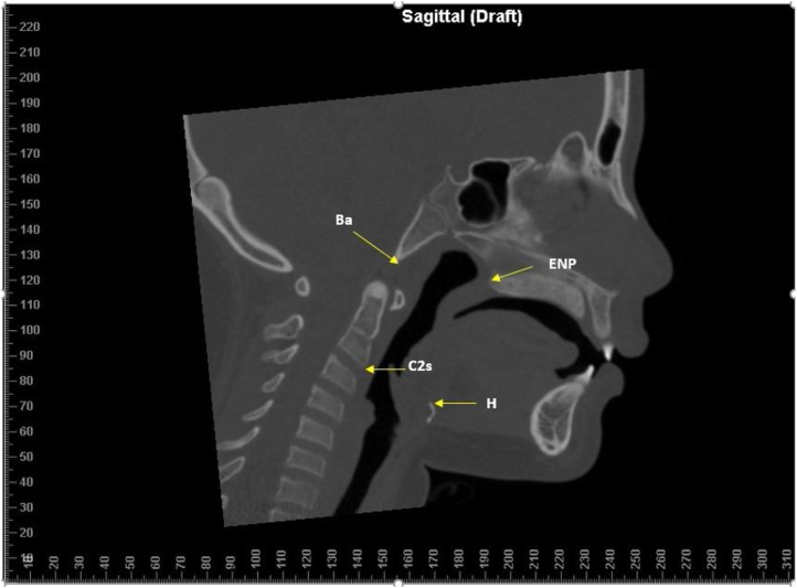
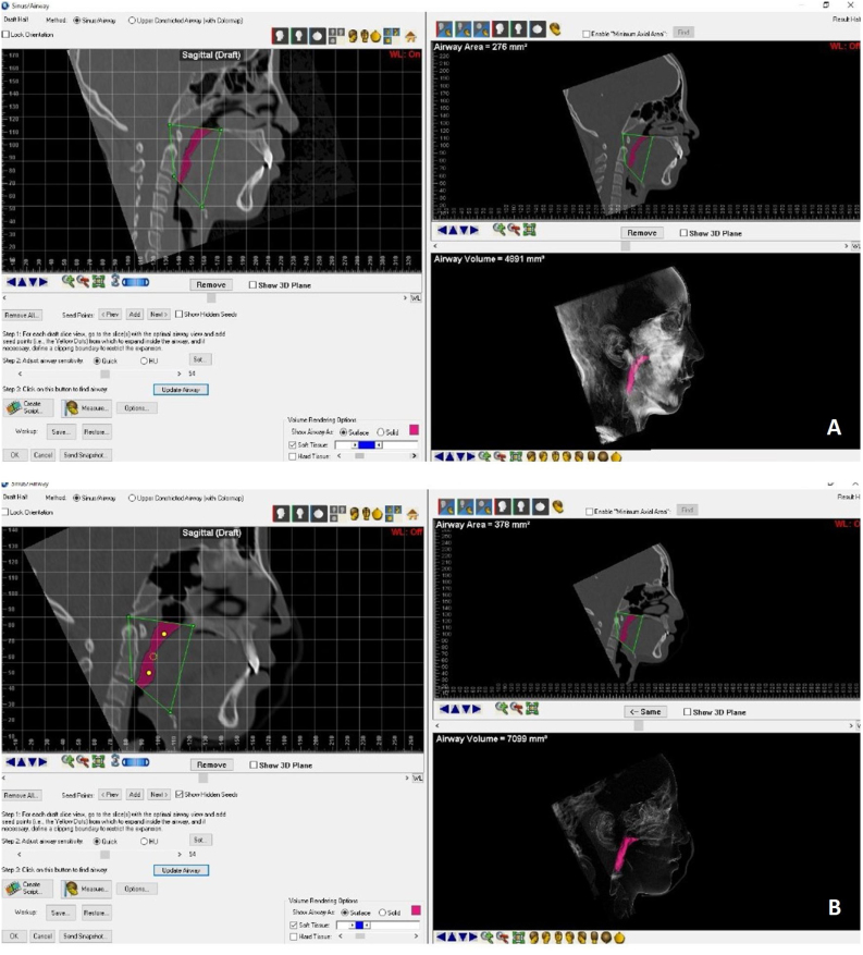
Twenty-four patients (11 with obstructive sleep apnea and 13 with residual snoring, mean age 10.0 (1.8), were enrolled in the study. Each patient underwent multislice computed tomography and nocturnal polysomnography before rapid maxillary expansion and after removal of maxillary expansion after six months. Airway regions were segmented, and volumes were computed.
The increase in oropharyngeal volume was significant in both groups. Oxygen saturation and apnea/hypopnea index were not statistically significant. No correlation was found between total airway volume, oxygen saturation, and apnea/hypopnea index changes between the time points examined.
This study showed that when rapid maxillary expansion is performed in individuals with sleep-disordered breathing, there were statistically significant differences in oropharyngeal volume between pre- and post-rapid maxillary expansion, but there was no correlation between oxygen saturation values and oropharyngeal volume increase.
The article is classified as Evidence Level 3 (Three).
本前瞻性临床研究旨在探讨上颌快速扩弓对上气道的影响,将多排螺旋 CT 测量的气道容积与睡眠呼吸暂停低通气指数和血氧饱和度的多导睡眠图评估结果相关联。
共纳入 24 名患者(11 名阻塞性睡眠呼吸暂停患者和 13 名残留性打鼾患者,平均年龄 10.0(1.8)岁)。每位患者在快速上颌扩张前和 6 个月后上颌扩张去除后均进行多层螺旋 CT 和夜间多导睡眠图检查。对气道区域进行分割并计算体积。
两组患者的口咽腔体积均有显著增加。氧饱和度和呼吸暂停/低通气指数无统计学意义。在检查的时间点之间,总气道体积、氧饱和度和呼吸暂停/低通气指数变化之间未发现相关性。
本研究表明,在患有睡眠呼吸障碍的个体中进行上颌快速扩弓时,扩弓前后口咽腔体积存在统计学显著差异,但氧饱和度值与口咽腔体积增加之间无相关性。
该文章被归类为证据水平 3(三级)。