Križman Manja, Švara Tanja, Šoba Barbara, Rataj Aleksandra Vergles
Institute of Food Safety, Feed and Environment, Veterinary Faculty, University of Ljubljana, Gerbičeva 60, 1000, Ljubljana, Slovenia.
Institute of Pathology, Wild Animals, Fish and Bees, Veterinary Faculty, University of Ljubljana, Gerbičeva 60, 1000, Ljubljana, Slovenia.
Int J Parasitol Parasites Wildl. 2022 Jun 22;18:221-224. doi: 10.1016/j.ijppaw.2022.06.004. eCollection 2022 Aug.
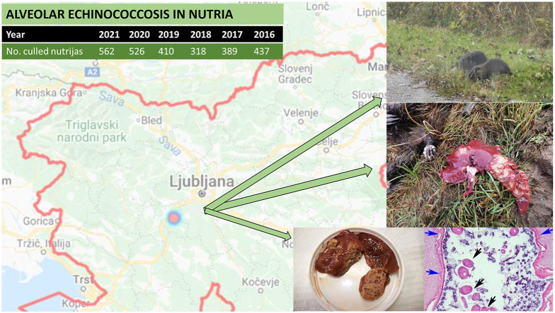
The present report describes a case of infection in nutria () culled in the central area of Slovenia. Post-mortem exam showed multiple cystic lesions in the liver. Gross examination, as well as parasitological and histopathological examinations, revealed numerous cysts of various sizes, filled with yellow clear fluid and displacing most of the liver parenchyma. The cyst lumina contained numerous protoscolices approximately 100 μm in diameter and calcareous corpuscles. The protoscolices had two visible suckers and a rostellum with birefringent hooks. The lesions were consistent with an cyst. Molecular analysis confirmed that the nutria was infected with . To our knowledge, this is the first report of echinococcosis in nutria in Slovenia that presents gross, parasitological, and histological lesions and the result of molecular analysis. Nutrias in Slovenia are dangerous invaders but can also be a relevant bioindicator of the presence of the parasite in the environment.
本报告描述了一例在斯洛文尼亚中部地区捕杀的海狸鼠感染病例。尸检显示肝脏有多个囊性病变。大体检查以及寄生虫学和组织病理学检查发现了许多大小各异的囊肿,囊肿内充满黄色清亮液体,占据了大部分肝实质。囊肿腔内含有许多直径约100μm的原头蚴和石灰小体。原头蚴有两个可见的吸盘和一个带有双折射钩的顶突。这些病变与棘球蚴囊肿一致。分子分析证实该海狸鼠感染了棘球绦虫。据我们所知,这是斯洛文尼亚海狸鼠棘球蚴病的首例报告,呈现了大体、寄生虫学和组织学病变以及分子分析结果。斯洛文尼亚的海狸鼠是危险的入侵物种,但也可能是环境中寄生虫存在的相关生物指示物。