Department of Molecular & Integrative Physiology, University of Michigan Medical School, Ann Arbor, MI, United States.
Department of Cell and Developmental Biology, University of Michigan Medical School, Ann Arbor, MI, United States.
Front Endocrinol (Lausanne). 2022 Jun 20;13:875865. doi: 10.3389/fendo.2022.875865. eCollection 2022.
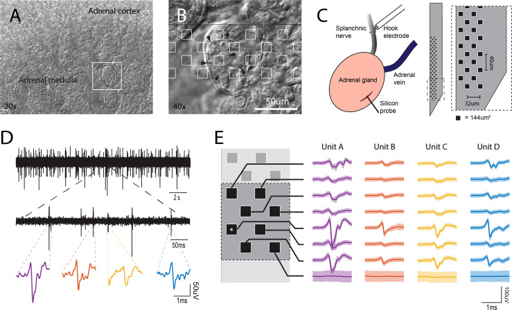
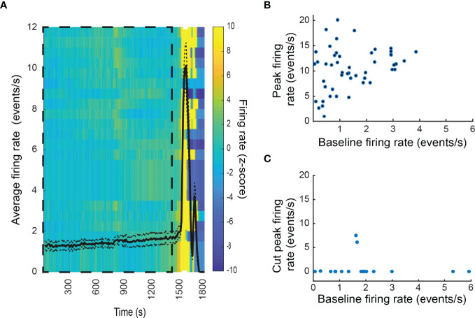
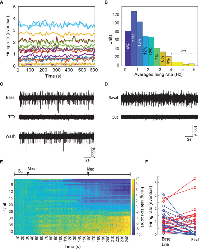
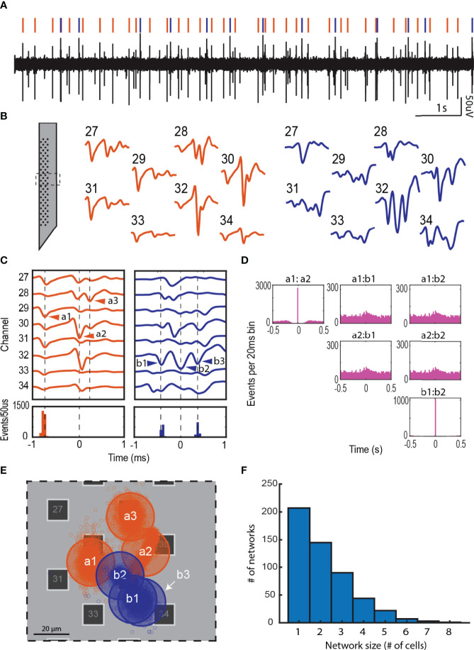
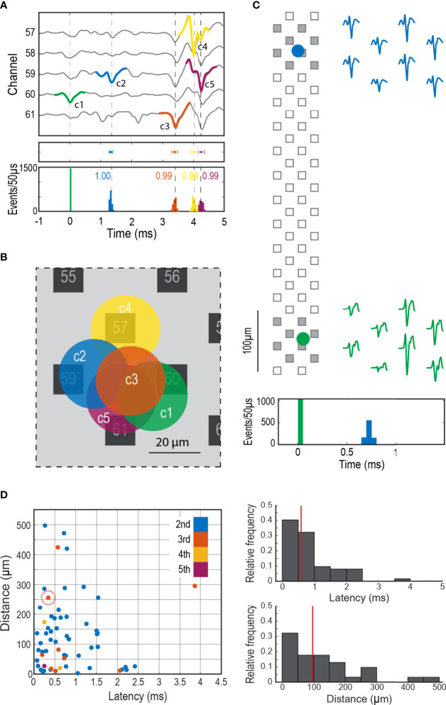
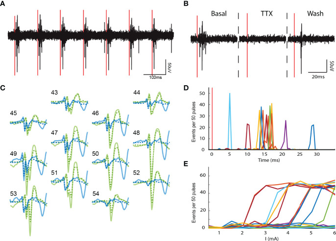
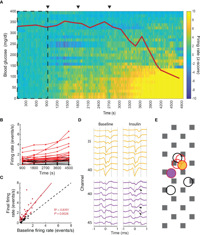
The adrenal medulla plays a critical role in mammalian homeostasis and the stress response. It is populated by clustered chromaffin cells that secrete epinephrine or norepinephrine along with peptides into the bloodstream affecting distant target organs. Despite been heavily studied, the central control of adrenal medulla and spatiotemporal responsiveness remains poorly understood. For this work, we continuously monitored the electrical activity of individual adrenomedullary chromaffin cells in the living anesthetized rat using multielectrode arrays. We measured the chromaffin cell activity under basal and physiological stress conditions and characterized the functional micro-architecture of the adrenal medulla. Under basal conditions, chromaffin cells fired action potentials with frequencies between ~0.2 and 4 Hz. Activity was almost completely driven by sympathetic inputs coming through the splanchnic nerve. Chromaffin cells were organized into independent local networks in which cells fired in a specific order, with latencies from hundreds of microseconds to a few milliseconds. Electrical stimulation of the splanchnic nerve evoked almost exactly the same spatiotemporal firing patterns that occurred spontaneously. Hypoglycemic stress, induced by insulin administration resulted in increased activity of a subset of the chromaffin cells. In contrast, respiratory arrest induced by lethal anesthesia resulted in an increase in the activity of virtually all chromaffin cells before cessation of all activity. These results suggest a stressor-specific activation of adrenomedullary chromaffin cell networks and revealed a surprisingly complex electrical organization that likely reflects the dynamic nature of the adrenal medulla's neuroendocrine output during basal conditions and during different types of physiological stress.
肾上腺髓质在哺乳动物的体内平衡和应激反应中起着关键作用。它由成群的嗜铬细胞组成,这些细胞会将肾上腺素或去甲肾上腺素以及肽类一起分泌到血液中,影响远处的靶器官。尽管已经进行了大量研究,但对肾上腺髓质的中枢控制和时空反应性仍了解甚少。在这项工作中,我们使用多电极阵列连续监测活体麻醉大鼠单个肾上腺髓质嗜铬细胞的电活动。我们在基础和生理应激条件下测量嗜铬细胞的活性,并对肾上腺髓质的功能微结构进行了特征描述。在基础条件下,嗜铬细胞以约 0.2 到 4 Hz 的频率发射动作电位。活动几乎完全由通过内脏神经传递的交感输入驱动。嗜铬细胞组织成独立的局部网络,其中细胞以特定的顺序发射,潜伏期从数百微秒到几毫秒不等。内脏神经的电刺激引发的时空发射模式几乎与自发发生的完全相同。胰岛素给药引起的低血糖应激导致一部分嗜铬细胞的活性增加。相比之下,致命麻醉引起的呼吸暂停导致几乎所有嗜铬细胞的活动增加,然后所有活动停止。这些结果表明,应激源特异性地激活了肾上腺髓质嗜铬细胞网络,并揭示了一种令人惊讶的复杂电组织,这可能反映了在基础条件下和不同类型的生理应激期间,肾上腺髓质神经内分泌输出的动态性质。