Achaiah Andrew, Lyon Paul, Fraser Emily, Saunders Peter, Hoyles Rachel, Benamore Rachel, Ho Ling-Pei
MRC Human Immunology Unit, Weatherall Institute of Molecular Medicine, University of Oxford, Oxford, UK.
Oxford Interstitial Lung Disease Service, Oxford University Hospitals NHS Foundation Trust, Oxford, UK.
ERJ Open Res. 2022 Jul 4;8(3). doi: 10.1183/23120541.00226-2022. eCollection 2022 Jul.
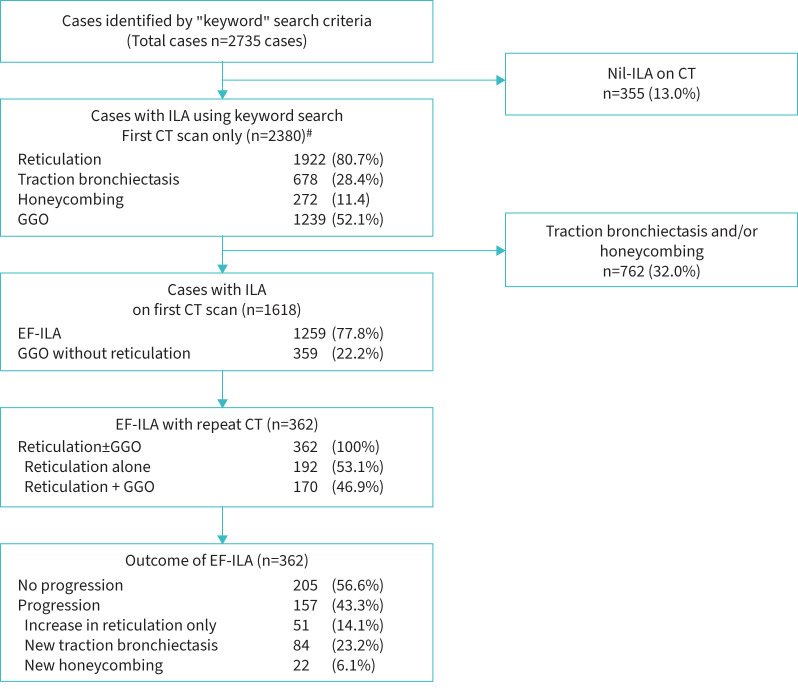
Interstitial lung abnormalities (ILA) are specific spatial patterns on computed tomography (CT) scan potentially compatible with early interstitial lung disease. A proportion will progress; management involves risk stratification and surveillance. Elevated blood monocyte levels have been shown to associate with progression of idiopathic pulmonary fibrosis. The aims of the present study were: 1) to estimate the proportion of "early fibrotic" (EF)-ILAs (reticular±ground-glass opacities, excluding traction bronchiectasis and honeycombing) on CT scans of patients attending all-indications thoracic CTs, and proportion demonstrating radiological progression; and 2) to explore association between peripheral blood leukocyte levels and ILA progression.
We analysed all thoracic CT reports in individuals aged 45-75 years performed between January 2015 and December 2020 in one large teaching hospital (Oxford, UK) to identify patient CT reports consistent with EF-ILA. CT-contemporaneous blood leukocyte counts were examined to explore contribution to progression and all-cause mortality, using multivariate Cox regression.
40 711 patients underwent thoracic CT imaging during this period. 1259 (3.1%) demonstrated the EF-ILA pattern (mean±sd age 65.4±7.32 years; 735 (47.8%) male). EF-ILA was significantly associated with all-cause mortality (hazard ratio 1.87, 95% CI 1.25-2.78; p=0.002). 362 cases underwent at least one follow-on CT. Radiological progression was observed in 157 (43.4%) cases: increase in reticulation n=51, new traction bronchiectasis n=84, honeycombing n=22. Monocyte count, neutrophil count, monocyte:lymphocyte ratio, neutrophil:lymphocyte ratio and "systemic inflammatory response index" were significantly associated with radiological progression.
3.1% of subjects requiring thoracic CT during a 6-year period demonstrated EF-ILA. Monocyte levels and blood leukocyte-derived indexes were associated with radiological progression and could indicate which patients may require closer follow-up.
间质性肺异常(ILA)是计算机断层扫描(CT)上特定的空间模式,可能与早期间质性肺疾病相符。一部分会进展;管理包括风险分层和监测。已显示血液单核细胞水平升高与特发性肺纤维化的进展相关。本研究的目的是:1)估计在因各种指征进行胸部CT检查的患者的CT扫描上“早期纤维化”(EF)-ILA(网状±磨玻璃影,不包括牵拉性支气管扩张和蜂窝状改变)的比例,以及显示放射学进展的比例;2)探讨外周血白细胞水平与ILA进展之间的关联。
我们分析了2015年1月至2020年12月在一家大型教学医院(英国牛津)对45-75岁个体进行的所有胸部CT报告,以识别与EF-ILA一致的患者CT报告。使用多变量Cox回归检查与CT同期的血液白细胞计数,以探讨其对进展和全因死亡率的影响。
在此期间,40711例患者接受了胸部CT成像。1259例(3.1%)表现出EF-ILA模式(平均±标准差年龄65.4±7.32岁;735例(47.8%)为男性)。EF-ILA与全因死亡率显著相关(风险比1.87,95%可信区间1.25-2.78;p=0.002)。362例患者接受了至少一次随访CT检查。157例(43.4%)观察到放射学进展:网状影增加n=51例,新出现的牵拉性支气管扩张n=84例,蜂窝状改变n=22例。单核细胞计数、中性粒细胞计数、单核细胞:淋巴细胞比值、中性粒细胞:淋巴细胞比值和“全身炎症反应指数”与放射学进展显著相关。
在6年期间需要进行胸部CT检查的患者中,3.1%表现出EF-ILA。单核细胞水平和血液白细胞衍生指标与放射学进展相关,可表明哪些患者可能需要更密切的随访。