• 文献检索
  • 文档翻译
  • 深度研究
  • 学术资讯
  • Suppr Zotero 插件Zotero 插件
  • 邀请有礼
  • 套餐&价格
  • 历史记录
应用&插件
Suppr Zotero 插件Zotero 插件浏览器插件Mac 客户端Windows 客户端微信小程序
定价
高级版会员购买积分包购买API积分包
服务
文献检索文档翻译深度研究API 文档MCP 服务
关于我们
关于 Suppr公司介绍联系我们用户协议隐私条款
关注我们

Suppr 超能文献

核心技术专利:CN118964589B侵权必究
粤ICP备2023148730 号-1Suppr @ 2026

文献检索

告别复杂PubMed语法,用中文像聊天一样搜索,搜遍4000万医学文献。AI智能推荐,让科研检索更轻松。

立即免费搜索

文件翻译

保留排版,准确专业,支持PDF/Word/PPT等文件格式,支持 12+语言互译。

免费翻译文档

深度研究

AI帮你快速写综述,25分钟生成高质量综述,智能提取关键信息,辅助科研写作。

立即免费体验

青光眼局限性血管楔形缺损的定量。

Quantification of localised vascular wedge-shaped defects in glaucoma.

机构信息

Department of Clinical Medicine, Macquarie University, Sydney, New South Wales, Australia.

Department of Ophthalmology, Flinders Health and Medical Research Institute, Flinders University, Adelaide, South Australia, Australia.

出版信息

Clin Exp Ophthalmol. 2022 Sep;50(7):724-735. doi: 10.1111/ceo.14134. Epub 2022 Jul 26.

DOI:10.1111/ceo.14134
PMID:35796092
原文链接:https://pmc.ncbi.nlm.nih.gov/articles/PMC9796144/
Abstract

BACKGROUND

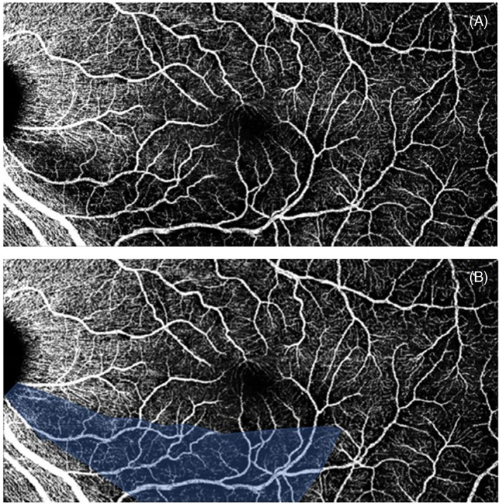
Vascular dysfunction plays a considerable role in glaucoma pathogenesis. Previous glaucoma case studies described localised wedge-shaped vascular defects, similar to retinal nerve fibre layer (RNFL) wedge defects. This study investigates the prevalence and quantification of this vessel loss, in relation to primary open angle glaucoma (POAG) parameters.

METHODS

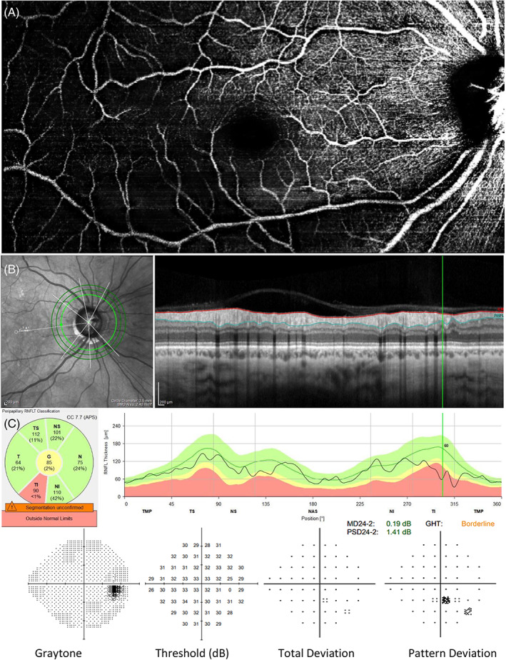
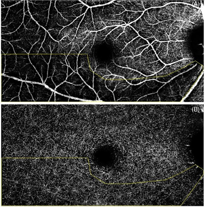
This study included 608 eyes (351 participants): 192 PROGRESSA study participants (342 eyes) with suspect, preperimetric or early manifest POAG, observed for vascular wedge defect presence (cohort one); an additional 114 individuals (cohort two-208 eyes) with POAG at various stages of progression for wedge characterisation; and 38 controls (56 eyes). Vascular wedge defects were observed using optical coherence tomography angiography (OCTA). Wedge parameters and vessel densities were quantified using ImageJ software. RNFL and ganglion cell layer inner plexiform layer (GCLIPL) from OCT scans, and mean deviation (Humphrey visual field 24-2) were also assessed.

RESULTS

Vascular wedge defects were found in 45/342 eyes (13.2%) in cohort one, in 41/208 eyes (19.7%) in cohort two and were not found in controls. Wedge defects were mostly inferotemporal (80%), and present at all disease stages. They were associated with visual field loss in the opposite hemisphere, thinner RNFL (p < 0.001), thinner GCLIPL (p = 0.003), and focal RNFL loss corresponding with the vascular defect region.

CONCLUSION

Vascular wedge defects are present at all POAG stages even before functional change and are strongly concordant with focal RNFL loss. Further research is needed to explore these defects in particular their temporal relationship with clinical measures of POAG.

摘要

背景

血管功能障碍在青光眼发病机制中起着重要作用。以前的青光眼病例研究描述了局部楔形血管缺陷,类似于视网膜神经纤维层 (RNFL) 楔形缺陷。本研究调查了这种血管损失的患病率和定量,与原发性开角型青光眼 (POAG) 参数有关。

方法

本研究包括 608 只眼(351 名参与者):192 名 PROGRESSA 研究参与者(342 只眼)患有可疑、前期或早期显性 POAG,观察血管楔形缺陷的存在(队列一);另外 114 名处于不同进展阶段的 POAG 患者(队列二-208 只眼)进行楔形特征描述;和 38 名对照者(56 只眼)。使用光学相干断层扫描血管造影术 (OCTA) 观察血管楔形缺陷。使用 ImageJ 软件定量楔形参数和血管密度。还评估了 OCT 扫描的 RNFL 和节细胞层内丛状层 (GCLIPL) 以及平均偏差 (Humphrey 视野 24-2)。

结果

在队列一的 342 只眼中发现了 45 只(13.2%)眼有血管楔形缺陷,在队列二的 208 只眼中发现了 41 只(19.7%)眼有血管楔形缺陷,对照组未发现。楔形缺陷主要位于下颞侧(80%),并存在于所有疾病阶段。它们与对侧半球的视野损失有关,与 RNFL 变薄(p<0.001)、GCLIPL 变薄(p=0.003)以及与血管缺陷区域相对应的局部 RNFL 损失有关。

结论

血管楔形缺陷存在于 POAG 的所有阶段,甚至在功能改变之前,并且与局部 RNFL 损失强烈一致。需要进一步研究这些缺陷,特别是它们与 POAG 的临床测量的时间关系。

https://cdn.ncbi.nlm.nih.gov/pmc/blobs/b7f7/9796144/af5738704cfa/CEO-50-724-g001.jpg
https://cdn.ncbi.nlm.nih.gov/pmc/blobs/b7f7/9796144/70ec8d9aeb1b/CEO-50-724-g003.jpg
https://cdn.ncbi.nlm.nih.gov/pmc/blobs/b7f7/9796144/eb082b4edaff/CEO-50-724-g002.jpg
https://cdn.ncbi.nlm.nih.gov/pmc/blobs/b7f7/9796144/af5738704cfa/CEO-50-724-g001.jpg
https://cdn.ncbi.nlm.nih.gov/pmc/blobs/b7f7/9796144/70ec8d9aeb1b/CEO-50-724-g003.jpg
https://cdn.ncbi.nlm.nih.gov/pmc/blobs/b7f7/9796144/eb082b4edaff/CEO-50-724-g002.jpg
https://cdn.ncbi.nlm.nih.gov/pmc/blobs/b7f7/9796144/af5738704cfa/CEO-50-724-g001.jpg

相似文献

1
Quantification of localised vascular wedge-shaped defects in glaucoma.青光眼局限性血管楔形缺损的定量。
Clin Exp Ophthalmol. 2022 Sep;50(7):724-735. doi: 10.1111/ceo.14134. Epub 2022 Jul 26.
2
Genetic risk of glaucoma is associated with vascular and retinal nerve fibre wedge defects.青光眼的遗传风险与血管及视网膜神经纤维楔形缺损相关。
Acta Ophthalmol. 2024 Mar;102(2):e185-e194. doi: 10.1111/aos.15775. Epub 2023 Oct 6.
3
Wedge Defects on Optical Coherence Tomography Angiography of the Peripapillary Retina in Glaucoma: Prevalence and Associated Clinical Factors.青光眼周边视网膜 OCT 血管造影的楔状缺损:患病率及相关临床因素。
J Glaucoma. 2022 Apr 1;31(4):242-249. doi: 10.1097/IJG.0000000000001991.
4
[Retinal vessel density in primary open-angle glaucoma with a hemifield defect].[原发性开角型青光眼伴半视野缺损的视网膜血管密度]
Zhonghua Yan Ke Za Zhi. 2021 Mar 11;57(3):201-206. doi: 10.3760/cma.j.cn112142-20201102-00734.
5
Localized wedge-shaped defects of retinal nerve fiber layer and disc hemorrhage in glaucoma.青光眼患者视网膜神经纤维层的局限性楔形缺损和视盘出血。
Ophthalmology. 1999 Sep;106(9):1762-7. doi: 10.1016/S0161-6420(99)90347-0.
6
Correlation between nerve fibre layer thickness measured with spectral domain OCT and visual field in patients with different stages of glaucoma.用谱域 OCT 测量的神经纤维层厚度与不同阶段青光眼患者视野的相关性。
Graefes Arch Clin Exp Ophthalmol. 2011 Apr;249(4):575-84. doi: 10.1007/s00417-010-1538-z. Epub 2010 Oct 15.
7
[The function-structure impairment pattern of optic nerves in primary open-angle glaucoma and normal-tension glaucoma].[原发性开角型青光眼和正常眼压性青光眼视神经的功能-结构损害模式]
Zhonghua Yan Ke Za Zhi. 2018 Nov 11;54(11):811-819. doi: 10.3760/cma.j.issn.0412-4081.2018.11.004.
8
Deep Retinal Layer Microvasculature Dropout Detected by the Optical Coherence Tomography Angiography in Glaucoma.光学相干断层扫描血管造影术检测青光眼患者视网膜深层微血管缺失
Ophthalmology. 2016 Dec;123(12):2509-2518. doi: 10.1016/j.ophtha.2016.09.002. Epub 2016 Oct 18.
9
[Analysis of morphologic changes of lamina cribrosa in primary open angle glaucoma using enhanced depth imaging optical coherence tomography].[使用增强深度成像光学相干断层扫描分析原发性开角型青光眼筛板的形态学变化]
Zhonghua Yan Ke Za Zhi. 2016 Jun 11;52(6):422-8. doi: 10.3760/cma.j.issn.0412-4081.2016.06.006.
10
Optical Coherence Tomography Angiography Vessel Density in Glaucomatous Eyes with Focal Lamina Cribrosa Defects.患有局限性筛板缺损的青光眼患者眼中光学相干断层扫描血管造影的血管密度
Ophthalmology. 2016 Nov;123(11):2309-2317. doi: 10.1016/j.ophtha.2016.07.023. Epub 2016 Aug 31.

引用本文的文献

1
Brain-Derived Neurotrophic Factor Val66Met is Associated with Variation in Cortical Structure in Healthy Aging Subjects.脑源性神经营养因子 Val66Met 与健康老年人皮质结构的变化有关。
Aging Dis. 2024 Oct 1;15(5):2315-2327. doi: 10.14336/AD.2024.0346.
2
Response of Diabetic Macular Edema to Anti-VEGF Medications Correlates with Improvement in Macular Vessel Architecture Measured with OCT Angiography.糖尿病性黄斑水肿对抗血管内皮生长因子药物的反应与光学相干断层扫描血管造影测量的黄斑血管结构改善相关。
Ophthalmol Sci. 2024 Jan 26;4(4):100478. doi: 10.1016/j.xops.2024.100478. eCollection 2024 Jul-Aug.
3
The Association of 4 Allele with Retinal Layer Thickness and Microvasculature in Older Adults: Optic Nerve Decline and Cognitive Change Study.

本文引用的文献

1
Diagnostic ability and sectoral structure-function relationship of circumpapillary and macular superficial vessel density in early glaucomatous eyes.早期青光眼眼中周边和黄斑浅层血管密度的诊断能力和节段性结构-功能关系。
Sci Rep. 2022 Apr 9;12(1):5991. doi: 10.1038/s41598-022-10033-1.
2
Optical coherence tomography angiography metrics predict normal tension glaucoma progression.光学相干断层扫描血管造影指标可预测正常眼压性青光眼的进展。
Acta Ophthalmol. 2022 Nov;100(7):e1455-e1462. doi: 10.1111/aos.15117. Epub 2022 Mar 8.
3
Optic Disc Hemorrhage Is Not Associated with Global Choroidal Vessel Loss, but Is Associated with Localized Choroidal Vessel Loss in Glaucoma.
4个等位基因与老年人视网膜层厚度和微血管系统的关联:视神经衰退与认知变化研究
J Clin Med. 2023 Sep 27;12(19):6219. doi: 10.3390/jcm12196219.
4
Retinal thickness and vascular parameters using optical coherence tomography in Alzheimer's disease: a meta-analysis.阿尔茨海默病中使用光学相干断层扫描测量视网膜厚度和血管参数的荟萃分析
Neural Regen Res. 2023 Nov;18(11):2504-2513. doi: 10.4103/1673-5374.371380.
视盘出血与全脉络膜血管丢失无关,但与青光眼患者的局部脉络膜血管丢失有关。
J Clin Med. 2022 Feb 18;11(4):1080. doi: 10.3390/jcm11041080.
4
Wedge Defects on Optical Coherence Tomography Angiography of the Peripapillary Retina in Glaucoma: Prevalence and Associated Clinical Factors.青光眼周边视网膜 OCT 血管造影的楔状缺损:患病率及相关临床因素。
J Glaucoma. 2022 Apr 1;31(4):242-249. doi: 10.1097/IJG.0000000000001991.
5
Multilayer Macula Vessel Density and Visual Field Progression in Glaucoma.青光眼的多层黄斑血管密度与视野进展。
Am J Ophthalmol. 2022 May;237:193-203. doi: 10.1016/j.ajo.2021.11.018. Epub 2021 Nov 19.
6
Multiple Systemic Vascular Risk Factors Are Associated With Low-Tension Glaucoma.多项系统性血管危险因素与低眼压性青光眼相关。
J Glaucoma. 2022 Jan 1;31(1):15-22. doi: 10.1097/IJG.0000000000001964.
7
Measurements of OCT Angiography Complement OCT for Diagnosing Early Primary Open-Angle Glaucoma.OCT 血管造影测量对早期原发性开角型青光眼的诊断有 OCT 的补充作用。
Ophthalmol Glaucoma. 2022 May-Jun;5(3):262-274. doi: 10.1016/j.ogla.2021.09.012. Epub 2021 Oct 9.
8
Role of hypertension as a risk factor for open-angle glaucoma: a systematic review and meta-analysis.高血压作为开角型青光眼危险因素的作用:一项系统评价和荟萃分析。
BMJ Open Ophthalmol. 2021 Sep 28;6(1):e000798. doi: 10.1136/bmjophth-2021-000798. eCollection 2021.
9
Rates of Circumpapillary Retinal Nerve Fiber Layer Thinning and Capillary Density Loss in Glaucomatous Eyes with Disc Hemorrhage.盘沿视网膜神经纤维层变薄和伴有盘出血的青光眼眼毛细血管密度丧失的比率。
Am J Ophthalmol. 2022 Mar;235:24-31. doi: 10.1016/j.ajo.2021.09.013. Epub 2021 Sep 26.
10
Superficial and Deep Macula Vessel Density in Healthy, Glaucoma Suspect, and Glaucoma Eyes.健康眼、青光眼疑似眼和青光眼眼中的浅层和深层黄斑血管密度。
J Glaucoma. 2021 Jun 1;30(6):e276-e284. doi: 10.1097/IJG.0000000000001860.