Department of Pure and Applied Chemistry, Technology and Innovation Centre, University of Strathclyde, 99 George Street, Glasgow G1 1RD, U.K.
The Defence Science and Technology Laboratory (Dstl), Porton Down, Salisbury SP4 0JQ, U.K.
ACS Appl Mater Interfaces. 2022 Jul 20;14(28):31613-31624. doi: 10.1021/acsami.2c05611. Epub 2022 Jul 8.
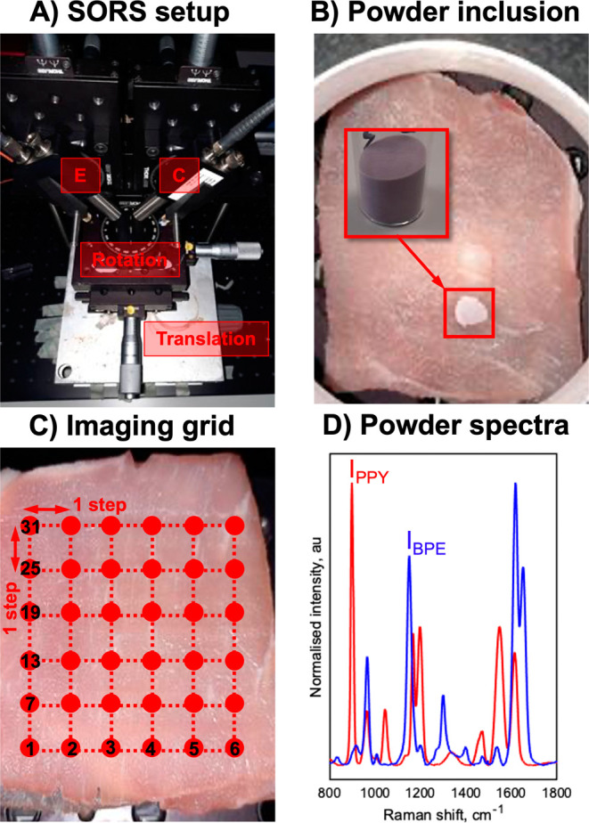
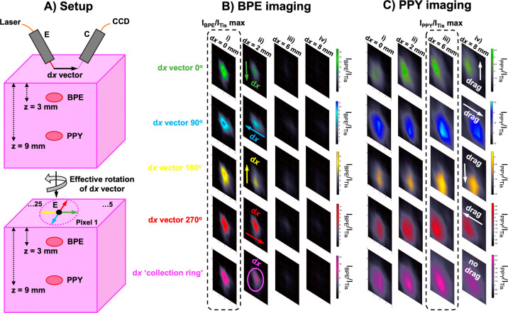
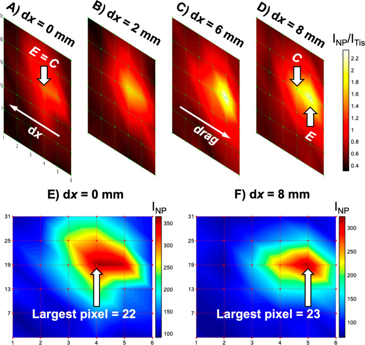
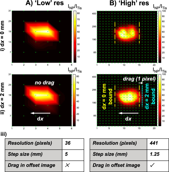
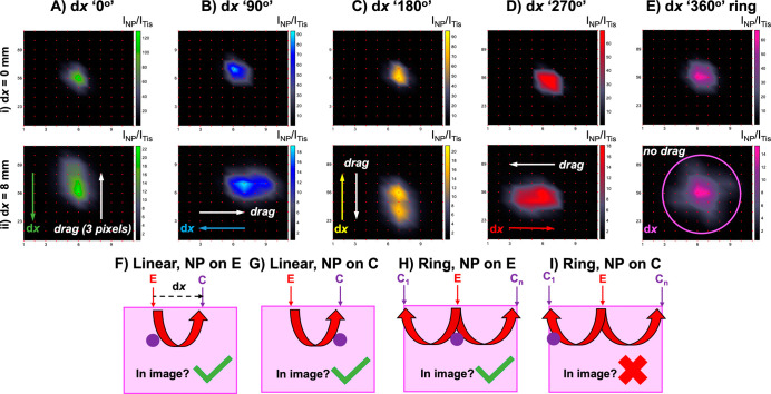
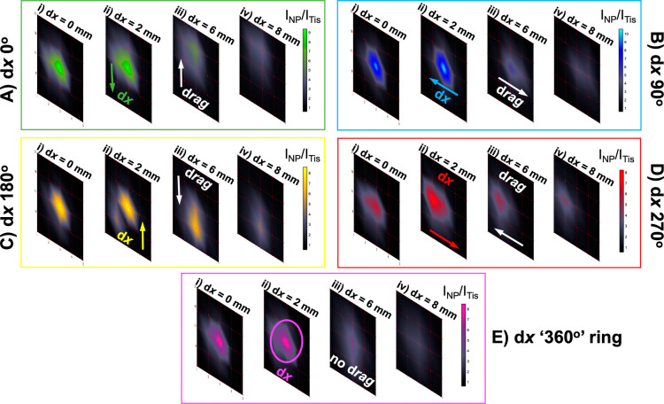
A fundamental question crucial to surface-enhanced spatially offset Raman spectroscopy (SESORS) imaging and implementing it in a clinical setting for in vivo diagnostic purposes is whether a SESORS image can be used to determine the exact location of an object within tissue? To address this question, multiple experimental factors pertaining to the optical setup in imaging experiments using an in-house-built point-collection-based spatially offset Raman spectroscopy (SORS) system were investigated to determine those critical to the three-dimensional (3D) positioning capability of SESORS. Here, we report the effects of the spatial offset magnitude and geometry on locating nanoparticles (NPs) mixed with silica powder as an imaging target through tissue and outline experimental techniques to allow for the correct interpretation of SESORS images to ascertain the correct location of NPs in the two-dimensional , -imaging plane at depth. More specifically, the effect of "linear offset-induced image drag" is presented, which refers to a spatial distortion in SESORS images caused by the magnitude and direction of the linear offset and highlight the need for an annular SORS collection geometry during imaging to neutralize these asymmetric effects. Additionally, building on these principles, the concept of "ratiometric SESORS imaging" is introduced for the location of buried inclusions in three dimensions. Together these principles are vital in developing a methodology for the location of surface-enhanced Raman scattering-active inclusions in three dimensions. This approach utilizes the relationship between the magnitude of the spatial offset, the probed depth, and ratiometric analysis of the NP and tissue Raman intensities to ultimately image and spatially discriminate between two distinct NP flavors buried at different depths within a 3D model for the first time. This research demonstrates how to accurately identify multiple objects at depth in tissue and their location using SESORS which addresses a key capability in moving SESORS closer to use in biomedical applications.
一个对于表面增强空间位移拉曼光谱(SESORS)成像至关重要的基本问题,也是将其应用于临床进行体内诊断的关键问题是,SESORS 图像是否可以用于确定组织内物体的精确位置?为了解决这个问题,本研究针对使用内部构建的基于点采集的空间位移拉曼光谱(SORS)系统进行成像实验的光学设置的多个实验因素进行了研究,以确定对 SESORS 三维(3D)定位能力至关重要的因素。在此,我们报告了空间位移幅度和几何形状对定位与二氧化硅粉末混合的纳米颗粒(NPs)的影响,这些 NPs 作为成像目标穿过组织,并概述了实验技术,以允许正确解释 SESORS 图像,从而确定 NPs 在二维成像平面中的正确位置在深度。更具体地说,提出了“线性偏移引起的图像拖曳”的影响,这是指 SESORS 图像中由于线性偏移的幅度和方向引起的空间变形,并强调在成像过程中需要采用环形 SORS 采集几何形状来中和这些不对称效应。此外,基于这些原理,引入了“比率 SESORS 成像”的概念,用于定位三维中的埋入式包裹体。这些原理对于开发一种用于定位三维中表面增强拉曼散射活性包裹体的方法至关重要。该方法利用空间偏移幅度、探测深度以及 NP 和组织拉曼强度的比率分析之间的关系,最终首次在三维模型中对埋藏在不同深度的两种不同 NP 风味进行成像和空间区分。这项研究展示了如何使用 SESORS 准确识别组织深处的多个物体及其位置,这是将 SESORS 更接近生物医学应用的关键能力之一。