• 文献检索
  • 文档翻译
  • 深度研究
  • 学术资讯
  • Suppr Zotero 插件Zotero 插件
  • 邀请有礼
  • 套餐&价格
  • 历史记录
应用&插件
Suppr Zotero 插件Zotero 插件浏览器插件Mac 客户端Windows 客户端微信小程序
定价
高级版会员购买积分包购买API积分包
服务
文献检索文档翻译深度研究API 文档MCP 服务
关于我们
关于 Suppr公司介绍联系我们用户协议隐私条款
关注我们

Suppr 超能文献

核心技术专利:CN118964589B侵权必究
粤ICP备2023148730 号-1Suppr @ 2026

文献检索

告别复杂PubMed语法,用中文像聊天一样搜索,搜遍4000万医学文献。AI智能推荐,让科研检索更轻松。

立即免费搜索

文件翻译

保留排版,准确专业,支持PDF/Word/PPT等文件格式,支持 12+语言互译。

免费翻译文档

深度研究

AI帮你快速写综述,25分钟生成高质量综述,智能提取关键信息,辅助科研写作。

立即免费体验

成人高弓足的评估与管理:一项叙述性综述

Evaluation and Management of Cavus Foot in Adults: A Narrative Review.

作者信息

Qin Boquan, Wu Shizhou, Zhang Hui

机构信息

Department of Orthopedics, Orthopedic Research Institute, West China Hospital, Sichuan University, Chengdu 610041, China.

出版信息

J Clin Med. 2022 Jun 26;11(13):3679. doi: 10.3390/jcm11133679.

DOI:10.3390/jcm11133679
PMID:35806964
原文链接:https://pmc.ncbi.nlm.nih.gov/articles/PMC9267353/
Abstract

OBJECTIVE

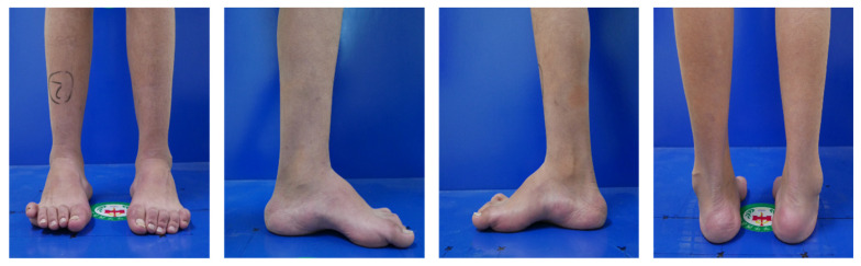
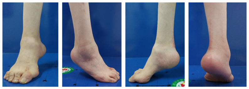
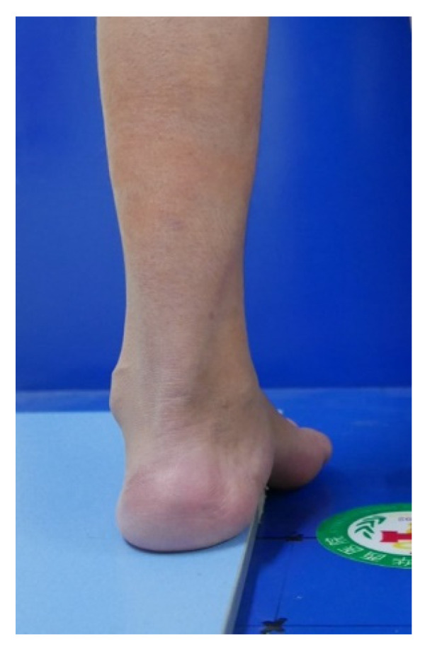
Cavus foot is a deformity defined by the abnormal elevation of the medial arch of the foot and is a common but challenging occurrence for foot and ankle surgeons. In this review, we mainly aim to provide a comprehensive evaluation of the treatment options available for cavus foot correction based on the current research and our experience and to highlight new technologies and future research directions.

METHODS

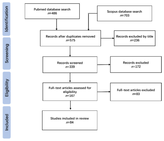
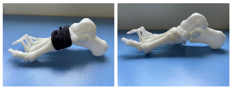
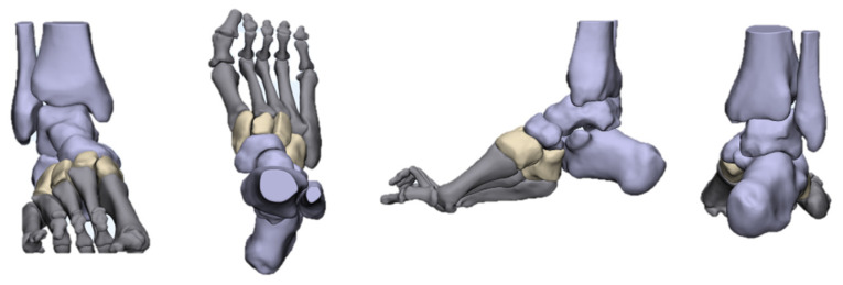
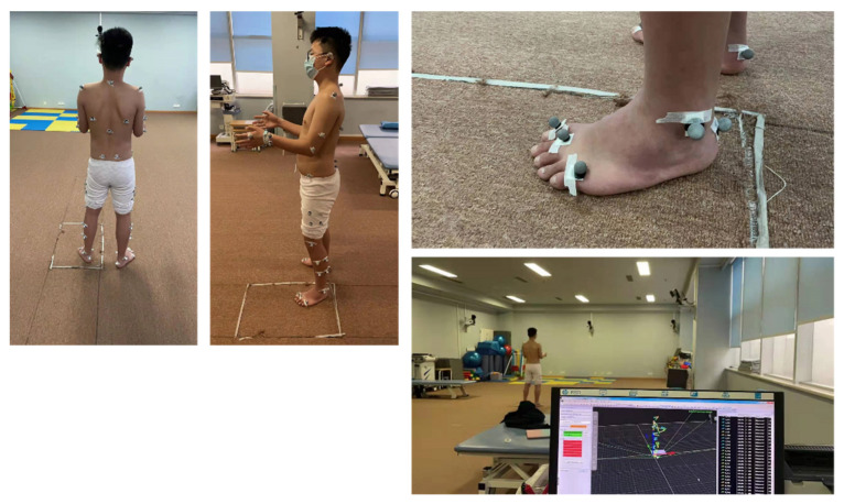
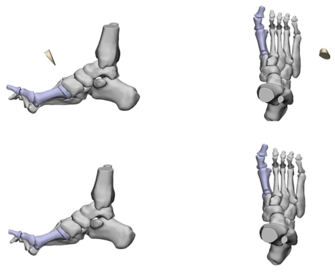
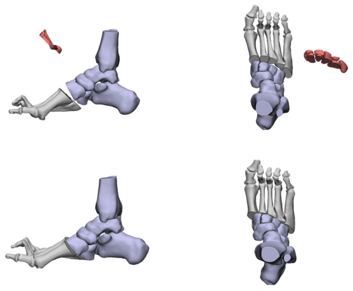
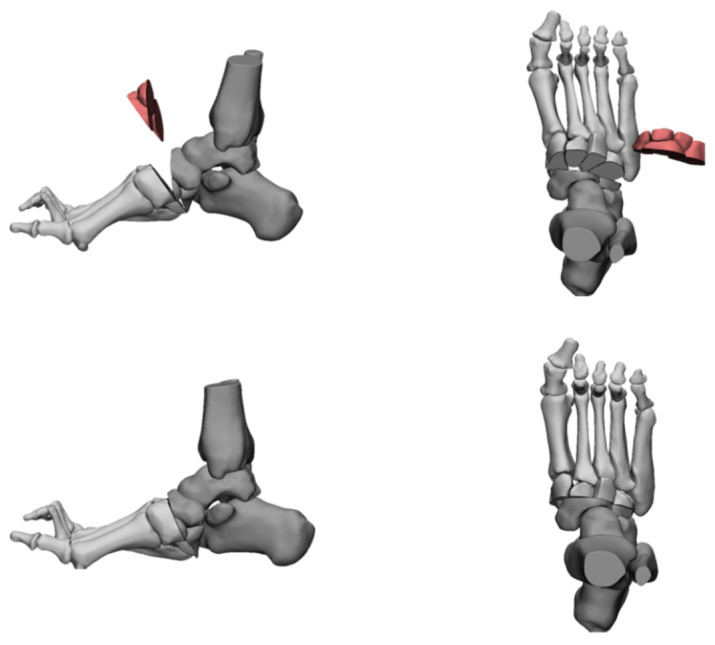
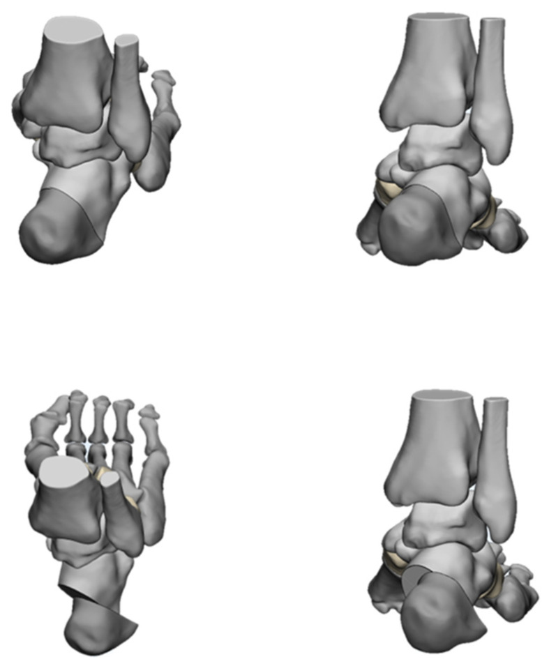
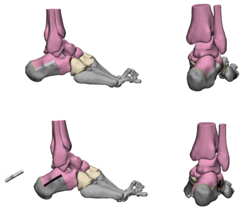
Searches on the PubMed and Scopus databases were conducted using the search terms cavus foot, CMT (Charcot-Marie-Tooth), tendon-transfer, osteotomy, and adult. The studies were screened according to the inclusion and exclusion criteria, and the correction of cavus foot was analyzed based on the current research and our own experience. At the same time, 3D models were used to simulate different surgical methods for cavus foot correction.

RESULTS

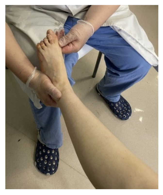
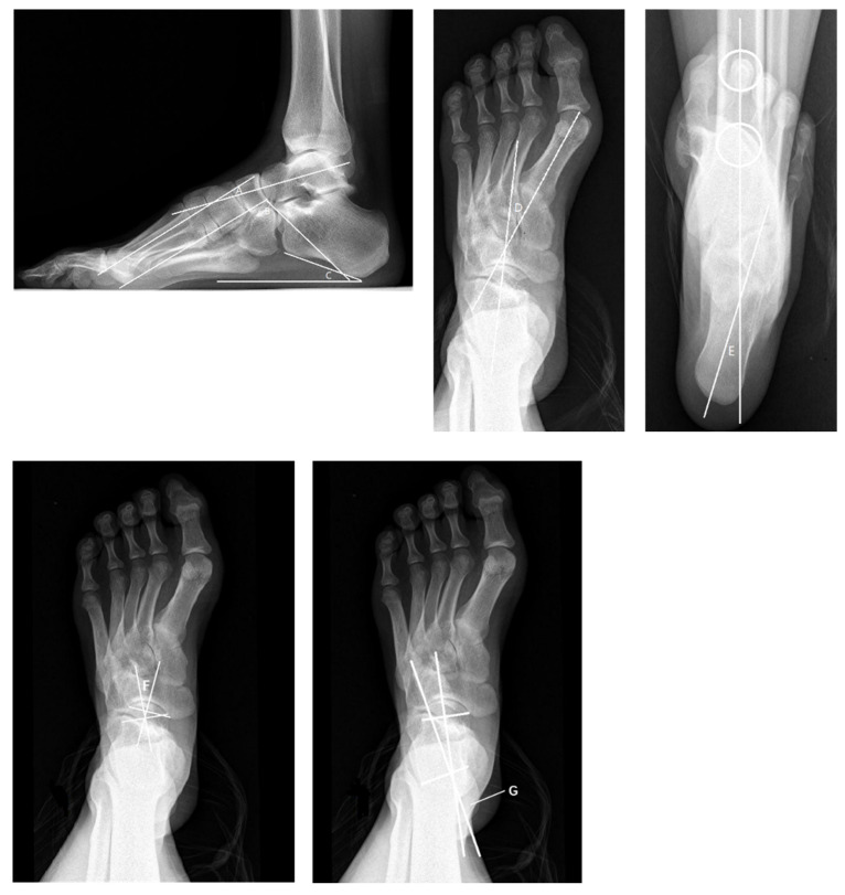
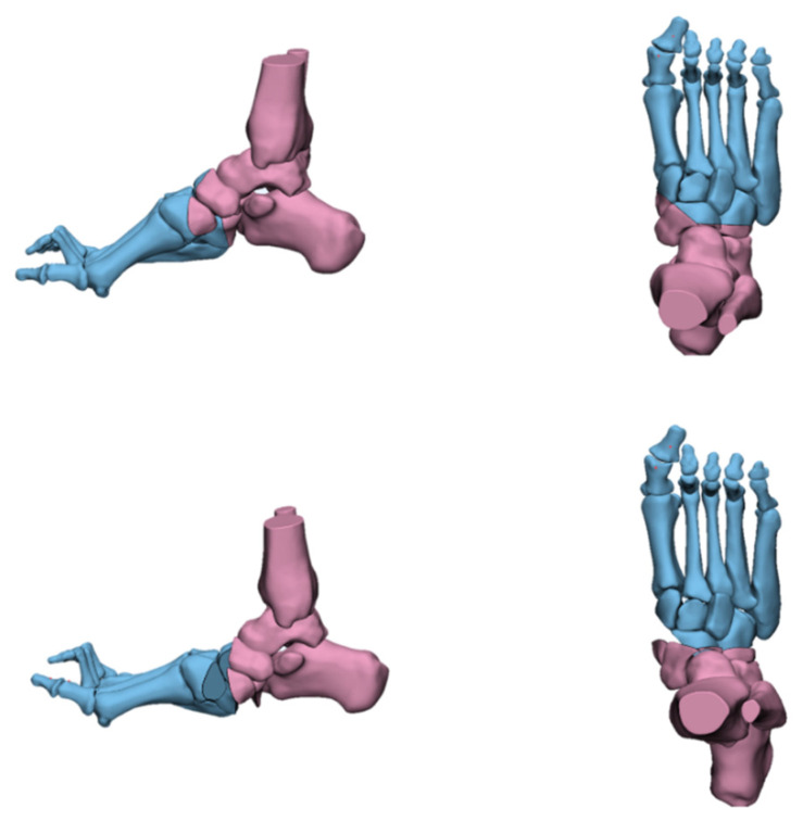
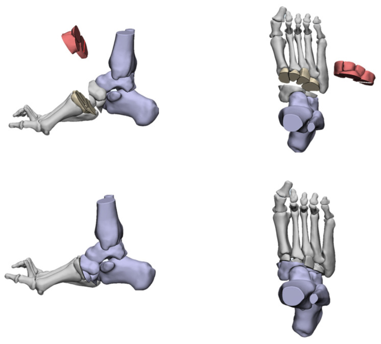
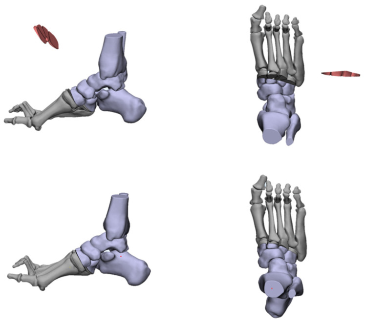
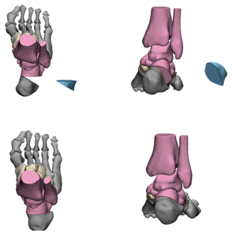
A total of 575 papers were identified and subsequently evaluated based on the title, abstract, and full text. A total of 84 articles were finally included in the review. The deformities involved in cavus foot are complex. Neuromuscular disorders are the main etiologies of cavus foot. Clinical evaluations including biomechanics, etiology, classification, pathophysiology and physical and radiological examinations should be conducted carefully in order to acquire a full understanding of cavus deformities. Soft-tissue release, tendon-transfer, and bony reconstruction are commonly used to correct cavus foot. Surgical plans need to be customized for different patients and usually involve a combination of multiple surgical procedures. A 3D simulation is helpful in that it allows us to gain a more intuitive understanding of various osteotomy methods.

CONCLUSION

The treatment of cavus foot requires us to make personalized operation plans according to different patients based on the comprehensive evaluation of their deformities. A combination of soft-tissue and bony procedures is required. Bony procedures are indispensable for cavus correction. With the promotion of digital orthopedics around the world, we can use computer technology to design and implement cavus foot operations in the future.

摘要

目的

高弓足是一种因足内侧弓异常抬高而定义的畸形,对于足踝外科医生来说是一种常见但具有挑战性的病症。在本综述中,我们主要旨在基于当前研究和我们的经验,对可用于高弓足矫正的治疗选择进行全面评估,并突出新技术和未来研究方向。

方法

使用搜索词“高弓足”“夏科-马里-图思病(CMT)”“肌腱转移”“截骨术”和“成人”在PubMed和Scopus数据库中进行检索。根据纳入和排除标准对研究进行筛选,并基于当前研究和我们自己的经验分析高弓足的矫正情况。同时,使用三维模型模拟高弓足矫正的不同手术方法。

结果

共识别出575篇论文,随后根据标题、摘要和全文进行评估。最终共有84篇文章纳入本综述。高弓足所涉及的畸形较为复杂。神经肌肉疾病是高弓足的主要病因。应仔细进行包括生物力学、病因、分类、病理生理学以及体格和影像学检查在内的临床评估,以便全面了解高弓畸形。软组织松解、肌腱转移和骨性重建是矫正高弓足常用的方法。手术方案需要针对不同患者进行定制,通常涉及多种手术的联合应用。三维模拟有助于我们更直观地了解各种截骨方法。

结论

高弓足的治疗需要我们在对患者畸形进行全面评估的基础上,根据不同患者制定个性化的手术方案。需要软组织和骨性手术相结合。骨性手术对于高弓矫正不可或缺。随着全球数字骨科的推广,未来我们可以利用计算机技术设计并实施高弓足手术。

https://cdn.ncbi.nlm.nih.gov/pmc/blobs/4088/9267353/2f59851cf18d/jcm-11-03679-g018.jpg
https://cdn.ncbi.nlm.nih.gov/pmc/blobs/4088/9267353/270e53cc6078/jcm-11-03679-g001.jpg
https://cdn.ncbi.nlm.nih.gov/pmc/blobs/4088/9267353/676453f09c2c/jcm-11-03679-g002.jpg
https://cdn.ncbi.nlm.nih.gov/pmc/blobs/4088/9267353/686906b3afae/jcm-11-03679-g003.jpg
https://cdn.ncbi.nlm.nih.gov/pmc/blobs/4088/9267353/9751e75c7aff/jcm-11-03679-g004.jpg
https://cdn.ncbi.nlm.nih.gov/pmc/blobs/4088/9267353/aeab7427243a/jcm-11-03679-g005.jpg
https://cdn.ncbi.nlm.nih.gov/pmc/blobs/4088/9267353/14acb5be585e/jcm-11-03679-g006.jpg
https://cdn.ncbi.nlm.nih.gov/pmc/blobs/4088/9267353/c76a70421448/jcm-11-03679-g007.jpg
https://cdn.ncbi.nlm.nih.gov/pmc/blobs/4088/9267353/c2bc86129546/jcm-11-03679-g008.jpg
https://cdn.ncbi.nlm.nih.gov/pmc/blobs/4088/9267353/badad9d88909/jcm-11-03679-g009.jpg
https://cdn.ncbi.nlm.nih.gov/pmc/blobs/4088/9267353/76deb4a47336/jcm-11-03679-g010.jpg
https://cdn.ncbi.nlm.nih.gov/pmc/blobs/4088/9267353/15a87cc81d90/jcm-11-03679-g011.jpg
https://cdn.ncbi.nlm.nih.gov/pmc/blobs/4088/9267353/865813c404c2/jcm-11-03679-g012.jpg
https://cdn.ncbi.nlm.nih.gov/pmc/blobs/4088/9267353/dbc814620a44/jcm-11-03679-g013.jpg
https://cdn.ncbi.nlm.nih.gov/pmc/blobs/4088/9267353/35ced4126539/jcm-11-03679-g014.jpg
https://cdn.ncbi.nlm.nih.gov/pmc/blobs/4088/9267353/fe79f274f653/jcm-11-03679-g015.jpg
https://cdn.ncbi.nlm.nih.gov/pmc/blobs/4088/9267353/ebf906a7d133/jcm-11-03679-g016.jpg
https://cdn.ncbi.nlm.nih.gov/pmc/blobs/4088/9267353/55510f431b17/jcm-11-03679-g017.jpg
https://cdn.ncbi.nlm.nih.gov/pmc/blobs/4088/9267353/2f59851cf18d/jcm-11-03679-g018.jpg

相似文献

1
Evaluation and Management of Cavus Foot in Adults: A Narrative Review.成人高弓足的评估与管理:一项叙述性综述
J Clin Med. 2022 Jun 26;11(13):3679. doi: 10.3390/jcm11133679.
2
Surgical treatment of cavus foot in Charcot-Marie-tooth disease: a review of twenty-four cases: AAOS exhibit selection.夏科-马里-图思病中高弓足的外科治疗:24例病例回顾:美国骨科学会展览入选报告
J Bone Joint Surg Am. 2015 Mar 18;97(6):e30. doi: 10.2106/JBJS.N.00794.
3
Long-term results of reconstruction for treatment of a flexible cavovarus foot in Charcot-Marie-Tooth disease.夏科-马里-图斯病中柔性高弓足治疗重建的长期结果。
J Bone Joint Surg Am. 2008 Dec;90(12):2631-42. doi: 10.2106/JBJS.G.01356.
4
Minimally Invasive Early Operative Treatment of Progressive Foot and Ankle Deformity Associated With Charcot-Marie-Tooth Disease.与夏科-马里-图思病相关的进行性足踝畸形的微创早期手术治疗
J Foot Ankle Surg. 2015 Jul-Aug;54(4):701-8. doi: 10.1053/j.jfas.2014.03.019. Epub 2014 Aug 15.
5
Pes cavus.高弓足
J Am Podiatr Med Assoc. 2005 Jan-Feb;95(1):42-52. doi: 10.7547/0950042.
6
Cavus foot deformity in children.儿童高弓足畸形
J Am Acad Orthop Surg. 2003 May-Jun;11(3):201-11. doi: 10.5435/00124635-200305000-00007.
7
The adult cavus foot.成人高弓足。
EFORT Open Rev. 2017 May 11;2(5):221-229. doi: 10.1302/2058-5241.2.160077. eCollection 2017 May.
8
Cavus foot reconstruction in 3 patients with Charcot-Marie-Tooth disease.3例夏科-马里-图思病患者的高弓足重建术。
J Foot Ankle Surg. 2009 Mar-Apr;48(2):116-24. doi: 10.1053/j.jfas.2008.11.013.
9
[Treatment for Patients with Charcot-Marie-Tooth Disease: Orthopaedic Aspects].[夏科-马里-图思病患者的治疗:骨科方面]
Brain Nerve. 2016 Jan;68(1):51-7. doi: 10.11477/mf.1416200345.
10
Treatment of the cavus foot. Deformity in the pediatric patient with Charcot-Marie-Tooth.高弓足的治疗。患有夏科-马里-图思病的儿科患者的畸形情况。
Foot Ankle Clin. 2000 Jun;5(2):305-15.

引用本文的文献

1
Nerve Diameter and DTI Parameters Maybe Potential Markers for Clinical Trial in Patients With Charcot-Marie-Tooth Disease Type 1A.神经直径和扩散张量成像参数可能是1A型遗传性运动感觉神经病患者临床试验的潜在标志物。
Eur J Neurol. 2025 Jun;32(6):e70220. doi: 10.1111/ene.70220.
2
Does the Sagittal Radiographic Morphology of Subtalar Joint Affect the Alignment of Foot?距下关节矢状面形态是否影响足的对线?
Orthop Surg. 2024 Jun;16(6):1269-1276. doi: 10.1111/os.14054. Epub 2024 Apr 15.

本文引用的文献

1
Cavovarus With a Twist: Midfoot Coronal and Axial Plane Rotational Deformity in Charcot-Marie-Tooth Disease.足内翻伴扭转畸形:Charcot-Marie-Tooth 病的中足冠状面和矢状面旋转畸形。
Foot Ankle Int. 2022 May;43(5):676-682. doi: 10.1177/10711007211064600. Epub 2022 Jan 15.
2
Abnormal Bone Morphology in Charcot-Marie-Tooth Disease.Charcot-Marie-Tooth 病中的异常骨骼形态。
Foot Ankle Int. 2022 Apr;43(4):576-581. doi: 10.1177/10711007211055460. Epub 2021 Dec 15.
3
Percutaneous plantar fasciotomy: radiological evolution of medial longitudinal arch and clinical results after one year.
经皮足底筋膜切开术:内侧纵弓的影像学演变及一年后的临床结果
Int Orthop. 2022 Apr;46(4):861-866. doi: 10.1007/s00264-021-05186-z. Epub 2021 Aug 18.
4
Neglected neurogenic clubfoot treated with Achilles tendon lengthening using Z-plasty, total talectomy, and tibiocalcaneal arthrodesis.采用Z形皮瓣延长跟腱、全距骨切除术及胫跟关节融合术治疗 neglected neurogenic clubfoot(未提及准确中文术语,可直译为“被忽视的神经源性马蹄内翻足” ) 。
Int J Surg Case Rep. 2021 Jul;84:106051. doi: 10.1016/j.ijscr.2021.106051. Epub 2021 Jun 2.
5
Tendon Transfers and Their Role in Cavus Foot Deformity.肌腱转移及其在马蹄内翻足畸形中的作用。
Clin Podiatr Med Surg. 2021 Jul;38(3):427-443. doi: 10.1016/j.cpm.2021.02.005.
6
Management of Midfoot Cavus.中足高弓的处理。
Clin Podiatr Med Surg. 2021 Jul;38(3):391-410. doi: 10.1016/j.cpm.2021.02.004.
7
Technique tip: 3D printing surgical guide for pes cavus midfoot osteotomy.技术提示:3D 打印手术导板治疗高弓中足骨切开术。
Foot Ankle Surg. 2022 Apr;28(3):371-377. doi: 10.1016/j.fas.2021.05.001. Epub 2021 May 7.
8
Identification of Gait-Cycle Phases for Prosthesis Control.用于假肢控制的步态周期阶段识别
Biomimetics (Basel). 2021 Mar 26;6(2):22. doi: 10.3390/biomimetics6020022.
9
Biomechanical effects of foot orthoses with and without a lateral bar in individuals with cavus feet during comfortable and fast walking.在舒适和快速行走时,具有和不具有侧条的足部矫形器对高弓足个体的生物力学影响。
PLoS One. 2021 Mar 17;16(3):e0248658. doi: 10.1371/journal.pone.0248658. eCollection 2021.
10
Complex foot deformities associated with lower limb deformities: a new therapeutic strategy for simultaneous correction using Ilizarov procedure together with osteotomy and soft tissue release.与下肢畸形相关的复杂足畸形:应用伊里扎洛夫技术联合截骨和软组织松解术同时矫正的新治疗策略。
J Orthop Surg Res. 2020 Oct 23;15(1):492. doi: 10.1186/s13018-020-02021-w.