• 文献检索
  • 文档翻译
  • 深度研究
  • 学术资讯
  • Suppr Zotero 插件Zotero 插件
  • 邀请有礼
  • 套餐&价格
  • 历史记录
应用&插件
Suppr Zotero 插件Zotero 插件浏览器插件Mac 客户端Windows 客户端微信小程序
定价
高级版会员购买积分包购买API积分包
服务
文献检索文档翻译深度研究API 文档MCP 服务
关于我们
关于 Suppr公司介绍联系我们用户协议隐私条款
关注我们

Suppr 超能文献

核心技术专利:CN118964589B侵权必究
粤ICP备2023148730 号-1Suppr @ 2026

文献检索

告别复杂PubMed语法,用中文像聊天一样搜索,搜遍4000万医学文献。AI智能推荐,让科研检索更轻松。

立即免费搜索

文件翻译

保留排版,准确专业,支持PDF/Word/PPT等文件格式,支持 12+语言互译。

免费翻译文档

深度研究

AI帮你快速写综述,25分钟生成高质量综述,智能提取关键信息,辅助科研写作。

立即免费体验

视网膜血管分形维数作为自动眼底图像分析系统的图像质量指标。

Fractal dimension of retinal vasculature as an image quality metric for automated fundus image analysis systems.

机构信息

College of Computer Science and Technology, Zhejiang University, 38 Zheda Road, Hangzhou, 310027, China.

Department of Electrical and Computer Engineering, University of Alberta, Edmonton, T6G 1H9, Canada.

出版信息

Sci Rep. 2022 Jul 13;12(1):11868. doi: 10.1038/s41598-022-16089-3.

DOI:10.1038/s41598-022-16089-3
PMID:35831401
原文链接:https://pmc.ncbi.nlm.nih.gov/articles/PMC9279448/
Abstract

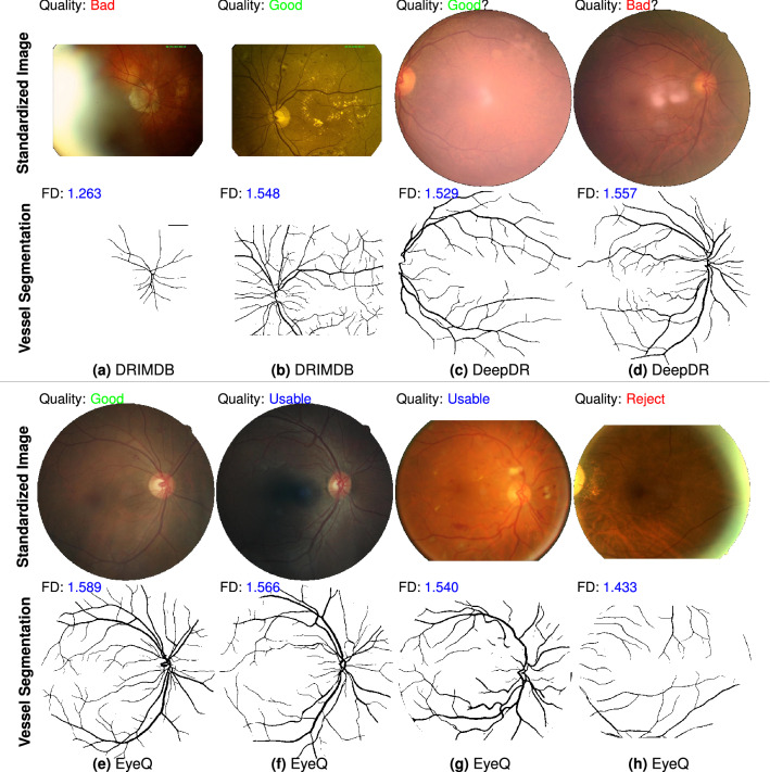
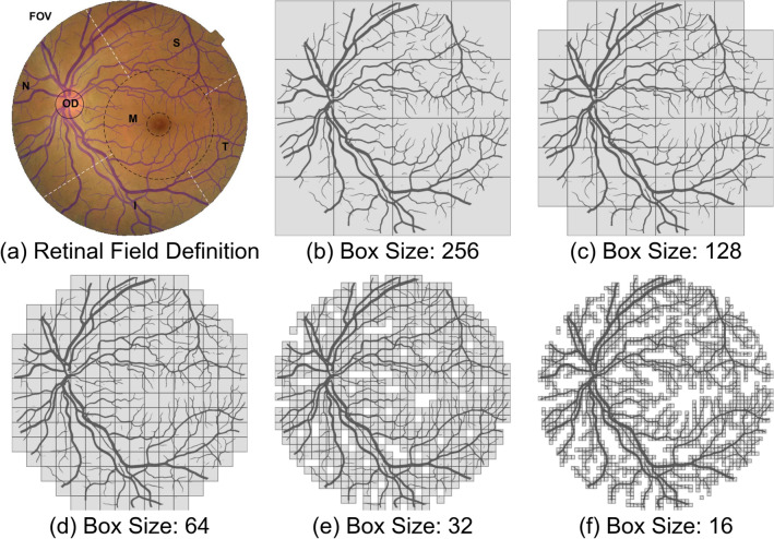
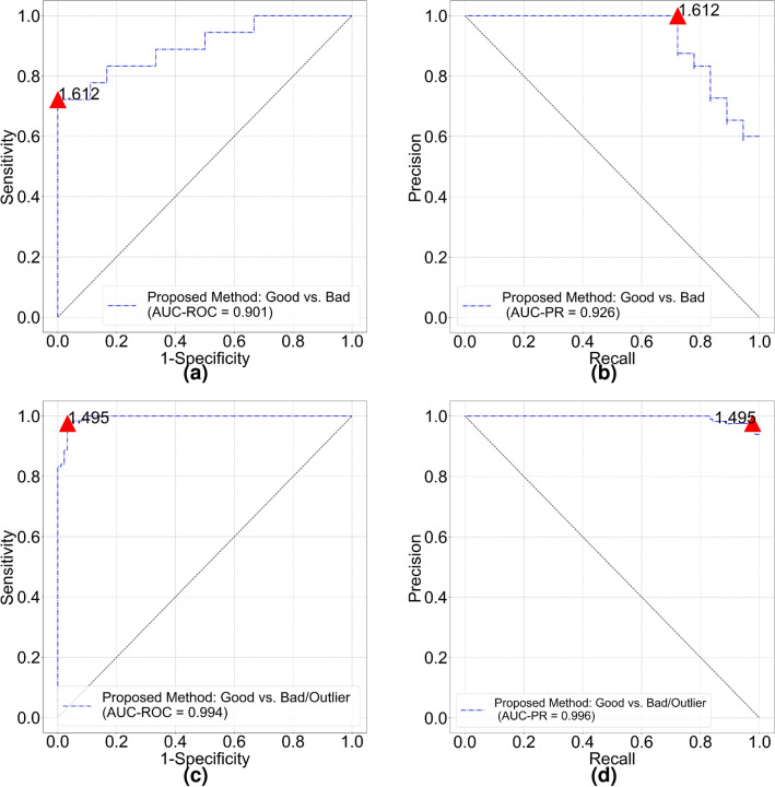
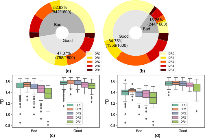
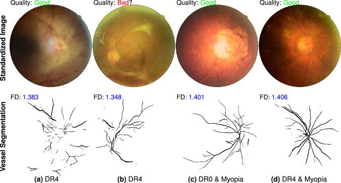
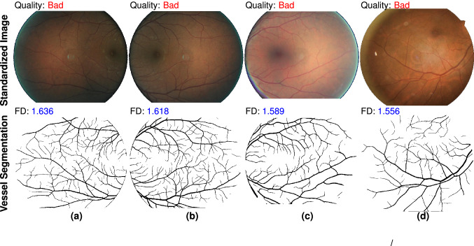
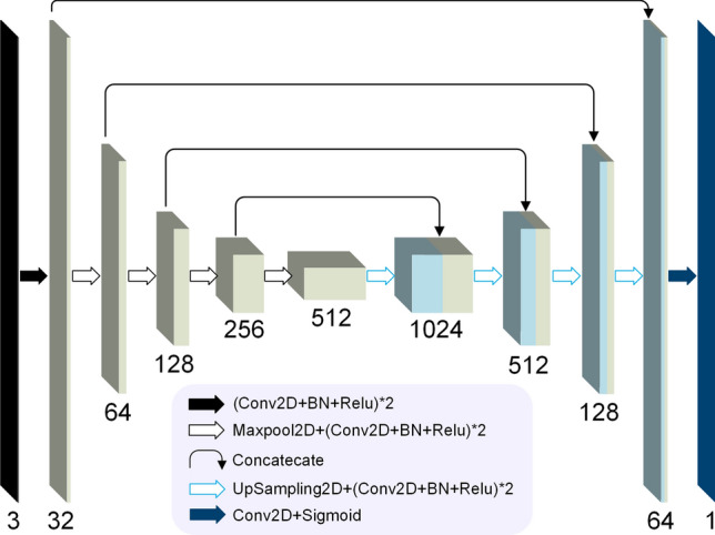
Automated fundus screening is becoming a significant programme of telemedicine in ophthalmology. Instant quality evaluation of uploaded retinal images could decrease unreliable diagnosis. In this work, we propose fractal dimension of retinal vasculature as an easy, effective and explainable indicator of retinal image quality. The pipeline of our approach is as follows: utilize image pre-processing technique to standardize input retinal images from possibly different sources to a uniform style; then, an improved deep learning empowered vessel segmentation model is employed to extract retinal vessels from the pre-processed images; finally, a box counting module is used to measure the fractal dimension of segmented vessel images. A small fractal threshold (could be a value between 1.45 and 1.50) indicates insufficient image quality. Our approach has been validated on 30,644 images from four public database.

摘要

自动眼底筛查正成为眼科远程医疗的一个重要项目。即时上传视网膜图像的质量评估可以减少不可靠的诊断。在这项工作中,我们提出视网膜血管的分形维数作为视网膜图像质量的一种简单、有效和可解释的指标。我们方法的流程如下:利用图像预处理技术将来自可能不同来源的输入视网膜图像标准化为统一的样式;然后,使用改进的深度学习赋能的血管分割模型从预处理图像中提取视网膜血管;最后,使用盒子计数模块测量分割血管图像的分形维数。较小的分形阈值(可能在 1.45 到 1.50 之间)表示图像质量不足。我们的方法已经在来自四个公共数据库的 30644 张图像上进行了验证。

https://cdn.ncbi.nlm.nih.gov/pmc/blobs/ac7c/9279448/7574688725d7/41598_2022_16089_Fig9_HTML.jpg
https://cdn.ncbi.nlm.nih.gov/pmc/blobs/ac7c/9279448/6e151bd0169d/41598_2022_16089_Fig1_HTML.jpg
https://cdn.ncbi.nlm.nih.gov/pmc/blobs/ac7c/9279448/b81e5185ecab/41598_2022_16089_Fig2_HTML.jpg
https://cdn.ncbi.nlm.nih.gov/pmc/blobs/ac7c/9279448/f899470b61f3/41598_2022_16089_Fig3_HTML.jpg
https://cdn.ncbi.nlm.nih.gov/pmc/blobs/ac7c/9279448/ec075751e8a2/41598_2022_16089_Fig4_HTML.jpg
https://cdn.ncbi.nlm.nih.gov/pmc/blobs/ac7c/9279448/3d8e24afc4d6/41598_2022_16089_Fig5_HTML.jpg
https://cdn.ncbi.nlm.nih.gov/pmc/blobs/ac7c/9279448/52b78abdc624/41598_2022_16089_Fig6_HTML.jpg
https://cdn.ncbi.nlm.nih.gov/pmc/blobs/ac7c/9279448/11a58fb20433/41598_2022_16089_Fig7_HTML.jpg
https://cdn.ncbi.nlm.nih.gov/pmc/blobs/ac7c/9279448/99b0fb909617/41598_2022_16089_Fig8_HTML.jpg
https://cdn.ncbi.nlm.nih.gov/pmc/blobs/ac7c/9279448/7574688725d7/41598_2022_16089_Fig9_HTML.jpg

相似文献

1
Fractal dimension of retinal vasculature as an image quality metric for automated fundus image analysis systems.视网膜血管分形维数作为自动眼底图像分析系统的图像质量指标。
Sci Rep. 2022 Jul 13;12(1):11868. doi: 10.1038/s41598-022-16089-3.
2
[Fractal analysis of retinal vascular tree: segmentation and estimation methods].[视网膜血管树的分形分析:分割与估计方法]
Arq Bras Oftalmol. 2007 May-Jun;70(3):413-22. doi: 10.1590/s0004-27492007000300006.
3
Robust methodology for fractal analysis of the retinal vasculature.视网膜血管的分形分析稳健方法。
IEEE Trans Med Imaging. 2011 Feb;30(2):243-50. doi: 10.1109/TMI.2010.2076322. Epub 2010 Sep 16.
4
DAVS-NET: Dense Aggregation Vessel Segmentation Network for retinal vasculature detection in fundus images.DAVS-NET:用于眼底图像中视网膜血管检测的密集聚合血管分割网络。
PLoS One. 2021 Dec 31;16(12):e0261698. doi: 10.1371/journal.pone.0261698. eCollection 2021.
5
Fractal analysis of normal retinal vascular network.正常视网膜血管网络的分形分析。
Oftalmologia. 2011;55(4):11-6.
6
Retinal Vasculature Fractal and Stroke Mortality.视网膜血管分形与中风死亡率。
Stroke. 2021 Apr;52(4):1276-1282. doi: 10.1161/STROKEAHA.120.031886. Epub 2021 Feb 22.
7
Fractal analysis of retinal vessel patterns in ophthalmically normal dogs.正常眼犬视网膜血管形态的分形分析
Vet Ophthalmol. 2008 Jan-Feb;11(1):2-6. doi: 10.1111/j.1463-5224.2007.00589.x.
8
SegR-Net: A deep learning framework with multi-scale feature fusion for robust retinal vessel segmentation.SegR-Net:一种具有多尺度特征融合的深度学习框架,用于稳健的视网膜血管分割。
Comput Biol Med. 2023 Sep;163:107132. doi: 10.1016/j.compbiomed.2023.107132. Epub 2023 Jun 10.
9
Quantification of Vessel Density in Retinal Optical Coherence Tomography Angiography Images Using Local Fractal Dimension.使用局部分形维数对视网膜光学相干断层扫描血管造影图像中的血管密度进行量化。
Invest Ophthalmol Vis Sci. 2016 Jan 1;57(1):246-52. doi: 10.1167/iovs.15-18287.
10
Network-based features for retinal fundus vessel structure analysis.基于网络的视网膜血管结构分析特征。
PLoS One. 2019 Jul 25;14(7):e0220132. doi: 10.1371/journal.pone.0220132. eCollection 2019.

引用本文的文献

1
Development and validation of a deep learning image quality feedback system for infant fundus photography.用于婴儿眼底摄影的深度学习图像质量反馈系统的开发与验证
Sci Rep. 2025 Jul 23;15(1):26852. doi: 10.1038/s41598-025-10859-5.
2
Evaluating the Impact of Retinal Vessel Segmentation Metrics on Retest Reliability in a Clinical Setting: A Comparative Analysis Using AutoMorph.评估视网膜血管分割指标对临床重测可靠性的影响:使用 AutoMorph 的对比分析
Invest Ophthalmol Vis Sci. 2024 Nov 4;65(13):24. doi: 10.1167/iovs.65.13.24.
3
Correlation between retinal vascular geometric parameters and pathologically diagnosed type 2 diabetic nephropathy.

本文引用的文献

1
The RETA Benchmark for Retinal Vascular Tree Analysis.RETA 视网膜血管树分析基准。
Sci Data. 2022 Jul 11;9(1):397. doi: 10.1038/s41597-022-01507-y.
2
DeepDRiD: Diabetic Retinopathy-Grading and Image Quality Estimation Challenge.深度糖尿病视网膜病变检测:糖尿病视网膜病变分级与图像质量评估挑战赛
Patterns (N Y). 2022 May 20;3(6):100512. doi: 10.1016/j.patter.2022.100512. eCollection 2022 Jun 10.
3
Joint optic disk and cup segmentation for glaucoma screening using a region-based deep learning network.基于区域的深度学习网络在青光眼筛查中的联合视盘和杯分割。
视网膜血管几何参数与病理诊断的2型糖尿病肾病之间的相关性。
Clin Kidney J. 2024 Jul 9;17(8):sfae204. doi: 10.1093/ckj/sfae204. eCollection 2024 Aug.
4
Evaluating imaging repeatability of fully self-service fundus photography within a community-based eye disease screening setting.评价基于社区眼病筛查环境下完全自助式眼底摄影的影像学重复性。
Biomed Eng Online. 2024 Mar 12;23(1):32. doi: 10.1186/s12938-024-01222-2.
5
Artificial Intelligence-based quantitative evaluation of retinal vascular parameters in thyroid-associated ophthalmopathy.基于人工智能的甲状腺相关性眼病视网膜血管参数定量评估
Endocrine. 2024 Jul;85(1):287-294. doi: 10.1007/s12020-023-03561-x. Epub 2024 Feb 3.
6
Pachychoroid neovasculopathy versus macular neovascularization in age-related macular degeneration with and without shallow irregular pigment epithelial detachment.年龄相关性黄斑变性伴或不伴浅性不规则色素上皮脱离的脉络膜增厚新生血管病变与黄斑新生血管
Sci Rep. 2023 Nov 9;13(1):19513. doi: 10.1038/s41598-023-46891-6.
7
Fractal nature of human gastrointestinal system: Exploring a new era.人类胃肠道系统的分形性质:探索新时代。
World J Gastroenterol. 2023 Jul 7;29(25):4036-4052. doi: 10.3748/wjg.v29.i25.4036.
8
Histologic Analysis of Idiopathic Pulmonary Fibrosis by Morphometric and Fractal Analysis.通过形态计量学和分形分析对特发性肺纤维化进行组织学分析。
Biomedicines. 2023 May 19;11(5):1483. doi: 10.3390/biomedicines11051483.
Eye (Lond). 2023 Apr;37(6):1080-1087. doi: 10.1038/s41433-022-02055-w. Epub 2022 Apr 18.
4
Feasibility and clinical utility of handheld fundus cameras for retinal imaging.手持式眼底相机进行视网膜成像的可行性和临床实用性。
Eye (Lond). 2023 Feb;37(2):274-279. doi: 10.1038/s41433-021-01926-y. Epub 2022 Jan 12.
5
An interpretable multiple-instance approach for the detection of referable diabetic retinopathy in fundus images.一种可解释的多实例方法,用于检测眼底图像中的可转诊糖尿病视网膜病变。
Sci Rep. 2021 Jul 12;11(1):14326. doi: 10.1038/s41598-021-93632-8.
6
Deep learning-based automated detection for diabetic retinopathy and diabetic macular oedema in retinal fundus photographs.基于深度学习的眼底图像糖尿病性视网膜病变和糖尿病性黄斑水肿自动检测。
Eye (Lond). 2022 Jul;36(7):1433-1441. doi: 10.1038/s41433-021-01552-8. Epub 2021 Jul 1.
7
A deep learning system for detecting diabetic retinopathy across the disease spectrum.一种用于在疾病谱中检测糖尿病性视网膜病变的深度学习系统。
Nat Commun. 2021 May 28;12(1):3242. doi: 10.1038/s41467-021-23458-5.
8
A review of machine learning methods for retinal blood vessel segmentation and artery/vein classification.机器学习方法在视网膜血管分割和动静脉分类中的研究进展综述。
Med Image Anal. 2021 Feb;68:101905. doi: 10.1016/j.media.2020.101905. Epub 2020 Nov 17.
9
Automatic fundus image quality assessment on a continuous scale.自动眼底图像质量连续评分。
Comput Biol Med. 2021 Feb;129:104114. doi: 10.1016/j.compbiomed.2020.104114. Epub 2020 Nov 12.
10
Pivotal trial of an autonomous AI-based diagnostic system for detection of diabetic retinopathy in primary care offices.在基层医疗诊所中用于检测糖尿病视网膜病变的基于人工智能的自主诊断系统的关键试验。
NPJ Digit Med. 2018 Aug 28;1:39. doi: 10.1038/s41746-018-0040-6. eCollection 2018.