Wang Yang-Kun, Zhou Jun-Ling, Meng Nian-Long, Zhu Chao-Ya, Wang Su-Nan, Chen Xiao-Dong
Department of Pathology, Foresea Life Insurance Guangzhou General Hospital, Guangzhou, 511300, People's Republic of China.
Shenzhen Nanshan District People's Hospital, Shenzhen, 518067, People's Republic of China.
Infect Drug Resist. 2022 Jul 7;15:3619-3629. doi: 10.2147/IDR.S355981. eCollection 2022.
To investigate the occurrence and development of gastric mucosal atrophy due to (Hp) infection and the accompanying histomorphological features.
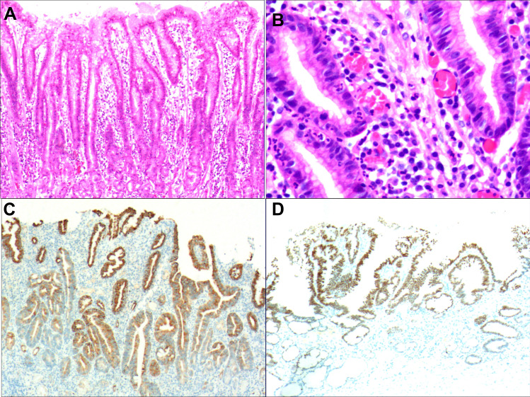
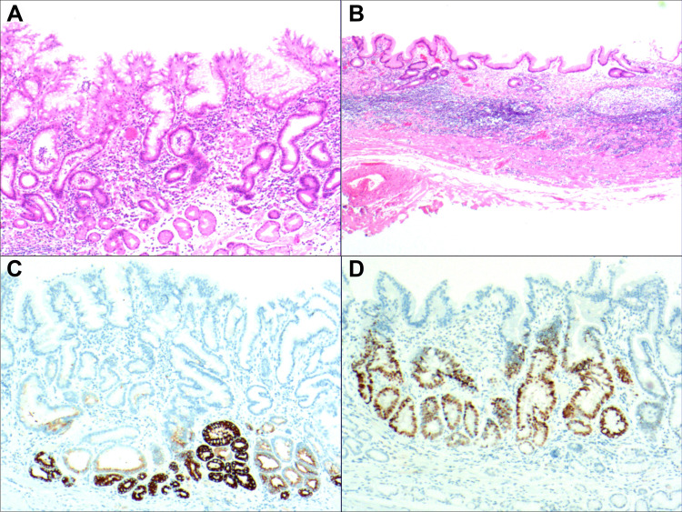
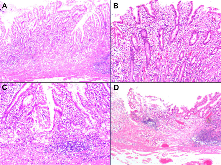
Detailed histological observations and immunohistochemical examinations were conducted via 197 endoscopic biopsies and endoscopic submucosal dissection specimens of gastric mucosal atrophic lesions with gastric Hp infection. Detailed observation was made of columnar cells in the proliferative region of the deep gastric pit and the isthmus of the gastric gland, as well as the upper part of the glandular cervix.
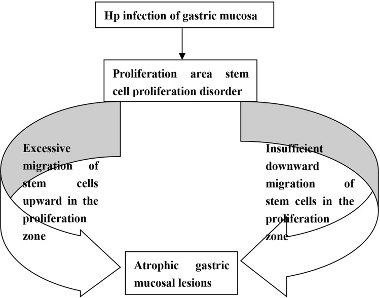
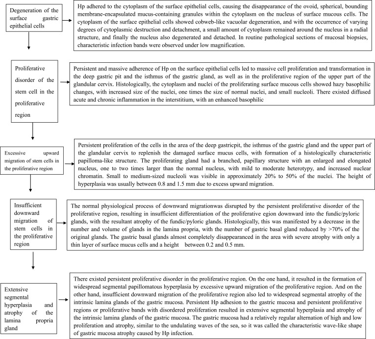
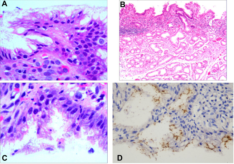
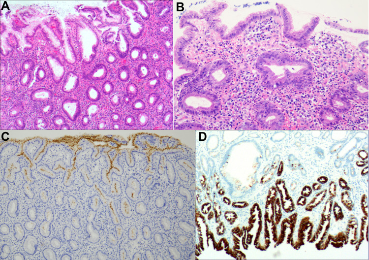
The infection of the gastric mucosa by Hp firstly led to the proliferative disorder of stem cells in the normal proliferative region of the gastric mucosa. This caused substantial propagation of cells in the proliferative region of the deep gastric pit and the isthmus of the gastric gland, as well as the upper part of the glandular cervix, as a means to replenish the damaged surface mucus cells. However, the propagation of stem cells in the proliferative region was insufficient for downward migration, and the normal physiological process of differentiation into fundic/pyloric gland cells was disrupted, resulting in glandular atrophy of the intrinsic layer of the gastric mucosa. Persistent Hp infection and disruption of stem cell proliferation in the proliferative region subsequently resulted in extensive segmental hyperplasia of the gastric mucosa and glandular atrophy of the lamina propria.
The occurrence, development, and histomorphological features of gastric mucosal atrophy due to gastric Hp infection provide a reliable pathological basis for precise treatment by clinicians and are of great significance for controlling the development of gastric cancer.
探讨幽门螺杆菌(Hp)感染所致胃黏膜萎缩的发生发展及伴随的组织形态学特征。
通过197例胃Hp感染的胃黏膜萎缩性病变的内镜活检及内镜黏膜下剥离标本进行详细的组织学观察和免疫组化检查。对胃小凹深部和胃腺峡部增殖区以及腺颈部上部的柱状细胞进行详细观察。
Hp感染胃黏膜首先导致胃黏膜正常增殖区干细胞增殖紊乱。这使得胃小凹深部和胃腺峡部以及腺颈部上部增殖区的细胞大量增殖,作为补充受损表面黏液细胞的一种方式。然而,增殖区干细胞的增殖不足以向下迁移,分化为胃底/幽门腺细胞的正常生理过程被破坏,导致胃黏膜固有层腺体萎缩。持续的Hp感染和增殖区干细胞增殖的破坏随后导致胃黏膜广泛的节段性增生和固有层腺体萎缩。
胃Hp感染所致胃黏膜萎缩的发生、发展及组织形态学特征为临床医生精准治疗提供了可靠的病理依据,对控制胃癌的发展具有重要意义。